Home > Press > Researchers demonstrate nanoscale X-ray imaging of bacterial cells
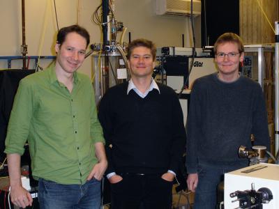 |
| Technische Universitaet Muenchen biophysicists (left to right) Dr. Pierre Thibault, Professor Dr. Franz Pfeiffer, and Martin Dierolf are co-authors of the PNAS paper, "Quantitative biological imaging by ptychographic X-ray diffraction microscopy." They collaborated with colleagues at the University of Goettingen and the Swiss Light Source. Credit: Andreas Battenberg, Technische Universitaet Muenchen. |
Abstract:
Sharper vision for new insights into biological questions, including DNA repair
Researchers demonstrate nanoscale X-ray imaging of bacterial cells
Germany | Posted on December 8th, 2009An ultra-high-resolution imaging technique using X-ray diffraction is a step closer to fulfilling its promise as a window on nanometer-scale structures in biological samples. In the Proceedings of the National Academy of Sciences, researchers report progress in applying an approach to "lensless" X-ray microscopy that they introduced one year ago. They have produced the first images, using this technique, of biological cells - specifically the intriguing polyextremophile Deinococcus radiourans. Better ability to see nanoscale structures in cells could yield important insights for evolutionary biology and biotechnology. In the case of D. radiourans, for example, it could help to settle questions about whether - or how - the structure of this organism's DNA-bearing nucleoid region accounts for its hardiness against ionizing radiation. Having demonstrated the resolution, reliability, and reproducibility of their technique, the researchers are now working to extend it to three-dimensional imaging of biological cells.
X-ray imaging is best known for its medical applications, such as traditional radiographs and CT scans. Yet the use of X-rays goes far beyond routine imaging. In particular, the very short wavelength of X-ray radiation allows various modes of microscopy that can reach the nanometer resolution. One of the main hurdles to high-resolution X-ray microscopy is the difficulty of producing high-quality X-ray lenses. To overcome these difficulties, so-called "lensless" microscopy methods have emerged in the last decade. A technique developed by researchers now in the biomedical physics group at Technische Universitaet Muenchen (TUM) has shown great promise for ultra-high resolution imaging of materials and life science samples.
This imaging technique, called ptychography, was first introduced in the 1970s for electron diffraction. It consists in measuring full far-field diffraction patterns as a small illumination is scanned on a sample. While its use in electron microscopy is still limited, ptychography has gained tremendous popularity in the X-ray imaging community in the last few years, thanks to the development by Franz Pfeiffer, now chair of the biomedical physics group at TUM, and his team. A critical step in the development of ptychography was published by the team one year ago in Science. The super-resolution capability of the imaging method was successfully demonstrated with a gold test structure.
Now a collaboration of the Pfeiffer group, together with researchers at University of Goettingen and at the Swiss Light Source (Villigen, Switzerland), has gone a step further and produced the first images of biological cells with the same technique.
These results, published in the Proceedings of the National Academy of Sciences, show that lensless X-ray imaging, in particular ptychography, can be used to obtain accurate maps of the electron density forming a biological sample. This type of quantitative measurement is extremely difficult with most other high-resolution techniques currently available. Moreover, biological samples are very fragile and nearly transparent to X-rays, making this type of accurate measurement even more challenging.
The Pfeiffer group is now moving beyond this success and looking into ways of improving the technique further. In particular, the team is aiming at the next milestone: three-dimensional imaging of biological samples.
This research is supported by the German Research Foundation (DFG), the Helmholtz Society, and the German Ministry of Education and Research.
Publications:
K. Giewekemeyer, P. Thibault, S. Kalbfleisch, A. Beerlink, C. M. Kewish, M. Dierolf, F. Pfeiffer, T. Salditt, Quantitative biological imaging by ptychographic x-ray diffraction microscopy, PNAS Early Edition, Proceedings of the National Academy of Sciences of the USA, Dec. 7-11, 2009. www.pnas.org/cgi/doi/10.1073/pnas.0905846107
P. Thibault, M. Dierolf, A. Menzel, O. Bunk, C. David, F. Pfeiffer, High-resolution scanning x-ray diffraction microscopy, Science 321, 379 - 381 (2008). www.sciencemag.org/cgi/content/abstract/321/5887/379
####
About Technische Universitaet Muenchen
Technische Universitaet Muenchen (TUM) is one of Germany's leading universities. It has roughly 440 professors, 6,500 academic and non-academic staff (including those at the university hospital "Rechts der Isar"), and 24,000 students. It focuses on the engineering sciences, natural sciences, life sciences, medicine, and economic sciences. After winning numerous awards, it was selected as an "Elite University" in 2006 by the Science Council (Wissenschaftsrat) and the German Research Foundation (DFG). The university's global network includes an outpost in Singapore. TUM is dedicated to the ideal of a top-level research based entrepreneurial university.
For more information, please click here
Contacts:
Andreas Battenberg
49-892-891-2890
Technische Universitaet Muenchen
Prof. Dr. Franz Pfeiffer
Chair for biomedical physics (E17)
Physics Department TUM
phone: +49 89 289 12552
Dr. Pierre Thibault
Physics Department TUM
phone: +49 89 289 14397
Copyright © Eurekalert
If you have a comment, please Contact us.Issuers of news releases, not 7th Wave, Inc. or Nanotechnology Now, are solely responsible for the accuracy of the content.
Bookmark:


| Related News Press |
News and information
![]() Quantum computer improves AI predictions April 17th, 2026
Quantum computer improves AI predictions April 17th, 2026
![]() Flexible sensor gains sensitivity under pressure April 17th, 2026
Flexible sensor gains sensitivity under pressure April 17th, 2026
![]() A reusable chip for particulate matter sensing April 17th, 2026
A reusable chip for particulate matter sensing April 17th, 2026
![]() Detecting vibrational quantum beating in the predissociation dynamics of SF6 using time-resolved photoelectron spectroscopy April 17th, 2026
Detecting vibrational quantum beating in the predissociation dynamics of SF6 using time-resolved photoelectron spectroscopy April 17th, 2026
Govt.-Legislation/Regulation/Funding/Policy
![]() Quantum computer improves AI predictions April 17th, 2026
Quantum computer improves AI predictions April 17th, 2026
![]() Metasurfaces smooth light to boost magnetic sensing precision January 30th, 2026
Metasurfaces smooth light to boost magnetic sensing precision January 30th, 2026
![]() New imaging approach transforms study of bacterial biofilms August 8th, 2025
New imaging approach transforms study of bacterial biofilms August 8th, 2025
Possible Futures
![]() A fundamentally new therapeutic approach to cystic fibrosis: Nanobody repairs cellular defect April 17th, 2026
A fundamentally new therapeutic approach to cystic fibrosis: Nanobody repairs cellular defect April 17th, 2026
![]() UC Irvine physicists discover method to reverse �quantum scrambling� : The work addresses the problem of information loss in quantum computing system April 17th, 2026
UC Irvine physicists discover method to reverse �quantum scrambling� : The work addresses the problem of information loss in quantum computing system April 17th, 2026
Nanomedicine
![]() A fundamentally new therapeutic approach to cystic fibrosis: Nanobody repairs cellular defect April 17th, 2026
A fundamentally new therapeutic approach to cystic fibrosis: Nanobody repairs cellular defect April 17th, 2026
![]() New molecular technology targets tumors and simultaneously silences two �undruggable� cancer genes August 8th, 2025
New molecular technology targets tumors and simultaneously silences two �undruggable� cancer genes August 8th, 2025
![]() New imaging approach transforms study of bacterial biofilms August 8th, 2025
New imaging approach transforms study of bacterial biofilms August 8th, 2025
![]() Electrifying results shed light on graphene foam as a potential material for lab grown cartilage June 6th, 2025
Electrifying results shed light on graphene foam as a potential material for lab grown cartilage June 6th, 2025
Announcements
![]() A fundamentally new therapeutic approach to cystic fibrosis: Nanobody repairs cellular defect April 17th, 2026
A fundamentally new therapeutic approach to cystic fibrosis: Nanobody repairs cellular defect April 17th, 2026
![]() UC Irvine physicists discover method to reverse �quantum scrambling� : The work addresses the problem of information loss in quantum computing system April 17th, 2026
UC Irvine physicists discover method to reverse �quantum scrambling� : The work addresses the problem of information loss in quantum computing system April 17th, 2026
Nanobiotechnology
![]() A fundamentally new therapeutic approach to cystic fibrosis: Nanobody repairs cellular defect April 17th, 2026
A fundamentally new therapeutic approach to cystic fibrosis: Nanobody repairs cellular defect April 17th, 2026
![]() New molecular technology targets tumors and simultaneously silences two �undruggable� cancer genes August 8th, 2025
New molecular technology targets tumors and simultaneously silences two �undruggable� cancer genes August 8th, 2025
![]() New imaging approach transforms study of bacterial biofilms August 8th, 2025
New imaging approach transforms study of bacterial biofilms August 8th, 2025
![]() Electrifying results shed light on graphene foam as a potential material for lab grown cartilage June 6th, 2025
Electrifying results shed light on graphene foam as a potential material for lab grown cartilage June 6th, 2025
Alliances/Trade associations/Partnerships/Distributorships
![]() Chicago Quantum Exchange welcomes six new partners highlighting quantum technology solutions, from Chicago and beyond September 23rd, 2022
Chicago Quantum Exchange welcomes six new partners highlighting quantum technology solutions, from Chicago and beyond September 23rd, 2022
![]() University of Illinois Chicago joins Brookhaven Lab's Quantum Center June 10th, 2022
University of Illinois Chicago joins Brookhaven Lab's Quantum Center June 10th, 2022
|
|
||
|
|
||
| The latest news from around the world, FREE | ||
|
|
||
|
|
||
| Premium Products | ||
|
|
||
|
Only the news you want to read!
Learn More |
||
|
|
||
|
Full-service, expert consulting
Learn More |
||
|
|
||