Home > Press > New optical imaging system could be deployed to find tiny tumors: Near-infrared technology pinpoints fluorescent probes deep within living tissue; may be used to detect cancer earlier
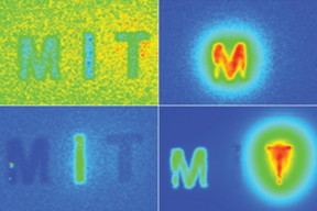 |
| MIT researchers have devised a way to simultaneously image in multiple wavelengths of near-infrared light, allowing them to determine the depth of particles emitting different wavelengths. Image courtesy of the researchers |
Abstract:
Many types of cancer could be more easily treated if they were detected at an earlier stage. MIT researchers have now developed an imaging system, named "DOLPHIN," which could enable them to find tiny tumors, as small as a couple of hundred cells, deep within the body.
New optical imaging system could be deployed to find tiny tumors: Near-infrared technology pinpoints fluorescent probes deep within living tissue; may be used to detect cancer earlier
Cambridge, MA | Posted on March 8th, 2019In a new study, the researchers used their imaging system, which relies on near-infrared light, to track a 0.1-millimeter fluorescent probe through the digestive tract of a living mouse. They also showed that they can detect a signal to a tissue depth of 8 centimeters, far deeper than any existing biomedical optical imaging technique.
The researchers hope to adapt their imaging technology for early diagnosis of ovarian and other cancers that are currently difficult to detect until late stages.
"We want to be able to find cancer much earlier," says Angela Belcher, the James Mason Crafts Professor of Biological Engineering and Materials Science at MIT and a member of the Koch Institute for Integrative Cancer Research, and the newly-appointed head of MIT's Department of Biological Engineering. "Our goal is to find tiny tumors, and do so in a noninvasive way."
Belcher is the senior author of the study, which appears in the March 7 issue of Scientific Reports. Xiangnan Dang, a former MIT postdoc, and Neelkanth Bardhan, a Mazumdar-Shaw International Oncology Fellow, are the lead authors of the study. Other authors include research scientists Jifa Qi and Ngozi Eze, former postdoc Li Gu, postdoc Ching-Wei Lin, graduate student Swati Kataria, and Paula Hammond, the David H. Koch Professor of Engineering, head of MIT's Department of Chemical Engineering, and a member of the Koch Institute.
Deeper imaging
Existing methods for imaging tumors all have limitations that prevent them from being useful for early cancer diagnosis. Most have a tradeoff between resolution and depth of imaging, and none of the optical imaging techniques can image deeper than about 3 centimeters into tissue. Commonly used scans such as X-ray computed tomography (CT) and magnetic resonance imaging (MRI) can image through the whole body; however, they can't reliably identify tumors until they reach about 1 centimeter in size.
Belcher's lab set out to develop new optical methods for cancer imaging several years ago, when they joined the Koch Institute. They wanted to develop technology that could image very small groups of cells deep within tissue and do so without any kind of radioactive labeling.
Near-infrared light, which has wavelengths from 900 to 1700 nanometers, is well-suited to tissue imaging because light with longer wavelengths doesn't scatter as much as when it strikes objects, which allows the light to penetrate deeper into the tissue. To take advantage of this, the researchers used an approach known as hyperspectral imaging, which enables simultaneous imaging in multiple wavelengths of light.
The researchers tested their system with a variety of near-infrared fluorescent light-emitting probes, mainly sodium yttrium fluoride nanoparticles that have rare earth elements such as erbium, holmium, or praseodymium added through a process called doping. Depending on the choice of the doping element, each of these particles emits near-infrared fluorescent light of different wavelengths.
Using algorithms that they developed, the researchers can analyze the data from the hyperspectral scan to identify the sources of fluorescent light of different wavelengths, which allows them to determine the location of a particular probe. By further analyzing light from narrower wavelength bands within the entire near-IR spectrum, the researchers can also determine the depth at which a probe is located. The researchers call their system "DOLPHIN", which stands for "Detection of Optically Luminescent Probes using Hyperspectral and diffuse Imaging in Near-infrared."
To demonstrate the potential usefulness of this system, the researchers tracked a 0.1-millimeter-sized cluster of fluorescent nanoparticles that was swallowed and then traveled through the digestive tract of a living mouse. These probes could be modified so that they target and fluorescently label specific cancer cells.
"In terms of practical applications, this technique would allow us to non-invasively track a 0.1-millimeter-sized fluorescently-labeled tumor, which is a cluster of about a few hundred cells. To our knowledge, no one has been able to do this previously using optical imaging techniques," Bardhan says.
Earlier detection
The researchers also demonstrated that they could inject fluorescent particles into the body of a mouse or a rat and then image through the entire animal, which requires imaging to a depth of about 4 centimeters, to determine where the particles ended up. And in tests with human tissue-mimics and animal tissue, they were able to locate the probes to a depth of up to 8 centimeters, depending on the type of tissue.
This kind of system could be used with any fluorescent probe that emits light in the near-infrared spectrum, including some that are already FDA-approved, the researchers say. The researchers are also working on adapting the imaging system so that it could reveal intrinsic differences in tissue contrast, including signatures of tumor cells, without any kind of fluorescent label.
In ongoing work, they are using a related version of this imaging system to try to detect ovarian tumors at an early stage. Ovarian cancer is usually diagnosed very late because there is no easy way to detect it when the tumors are still small.
"Ovarian cancer is a terrible disease, and it gets diagnosed so late because the symptoms are so nondescript," Belcher says. "We want a way to follow recurrence of the tumors, and eventually a way to find and follow early tumors when they first go down the path to cancer or metastasis. This is one of the first steps along the way in terms of developing this technology."
The researchers have also begun working on adapting this type of imaging to detect other types of cancer such as pancreatic cancer, brain cancer, and melanoma.
###
The research was funded by the Koch Institute Frontier Research Program, the Marble Center for Cancer Nanomedicine, the Koch Institute Support (core) Grant from the National Cancer Institute, the NCI Center for Center for Cancer Nanotechnology Excellence, and the Bridge Project.
####
For more information, please click here
Contacts:
Sarah McDonnell
617-253-8923
Copyright © Massachusetts Institute of Technology
If you have a comment, please Contact us.Issuers of news releases, not 7th Wave, Inc. or Nanotechnology Now, are solely responsible for the accuracy of the content.
Bookmark:


| Related News Press |
News and information
![]() Quantum computer improves AI predictions April 17th, 2026
Quantum computer improves AI predictions April 17th, 2026
![]() Flexible sensor gains sensitivity under pressure April 17th, 2026
Flexible sensor gains sensitivity under pressure April 17th, 2026
![]() A reusable chip for particulate matter sensing April 17th, 2026
A reusable chip for particulate matter sensing April 17th, 2026
![]() Detecting vibrational quantum beating in the predissociation dynamics of SF6 using time-resolved photoelectron spectroscopy April 17th, 2026
Detecting vibrational quantum beating in the predissociation dynamics of SF6 using time-resolved photoelectron spectroscopy April 17th, 2026
Imaging
![]() Simple algorithm paired with standard imaging tool could predict failure in lithium metal batteries August 8th, 2025
Simple algorithm paired with standard imaging tool could predict failure in lithium metal batteries August 8th, 2025
Govt.-Legislation/Regulation/Funding/Policy
![]() Quantum computer improves AI predictions April 17th, 2026
Quantum computer improves AI predictions April 17th, 2026
![]() Metasurfaces smooth light to boost magnetic sensing precision January 30th, 2026
Metasurfaces smooth light to boost magnetic sensing precision January 30th, 2026
![]() New imaging approach transforms study of bacterial biofilms August 8th, 2025
New imaging approach transforms study of bacterial biofilms August 8th, 2025
Possible Futures
![]() A fundamentally new therapeutic approach to cystic fibrosis: Nanobody repairs cellular defect April 17th, 2026
A fundamentally new therapeutic approach to cystic fibrosis: Nanobody repairs cellular defect April 17th, 2026
![]() UC Irvine physicists discover method to reverse �quantum scrambling� : The work addresses the problem of information loss in quantum computing system April 17th, 2026
UC Irvine physicists discover method to reverse �quantum scrambling� : The work addresses the problem of information loss in quantum computing system April 17th, 2026
Nanomedicine
![]() A fundamentally new therapeutic approach to cystic fibrosis: Nanobody repairs cellular defect April 17th, 2026
A fundamentally new therapeutic approach to cystic fibrosis: Nanobody repairs cellular defect April 17th, 2026
![]() New molecular technology targets tumors and simultaneously silences two �undruggable� cancer genes August 8th, 2025
New molecular technology targets tumors and simultaneously silences two �undruggable� cancer genes August 8th, 2025
![]() New imaging approach transforms study of bacterial biofilms August 8th, 2025
New imaging approach transforms study of bacterial biofilms August 8th, 2025
![]() Electrifying results shed light on graphene foam as a potential material for lab grown cartilage June 6th, 2025
Electrifying results shed light on graphene foam as a potential material for lab grown cartilage June 6th, 2025
Discoveries
![]() Quantum computer improves AI predictions April 17th, 2026
Quantum computer improves AI predictions April 17th, 2026
![]() Flexible sensor gains sensitivity under pressure April 17th, 2026
Flexible sensor gains sensitivity under pressure April 17th, 2026
![]() A reusable chip for particulate matter sensing April 17th, 2026
A reusable chip for particulate matter sensing April 17th, 2026
![]() Detecting vibrational quantum beating in the predissociation dynamics of SF6 using time-resolved photoelectron spectroscopy April 17th, 2026
Detecting vibrational quantum beating in the predissociation dynamics of SF6 using time-resolved photoelectron spectroscopy April 17th, 2026
Announcements
![]() A fundamentally new therapeutic approach to cystic fibrosis: Nanobody repairs cellular defect April 17th, 2026
A fundamentally new therapeutic approach to cystic fibrosis: Nanobody repairs cellular defect April 17th, 2026
![]() UC Irvine physicists discover method to reverse �quantum scrambling� : The work addresses the problem of information loss in quantum computing system April 17th, 2026
UC Irvine physicists discover method to reverse �quantum scrambling� : The work addresses the problem of information loss in quantum computing system April 17th, 2026
Interviews/Book Reviews/Essays/Reports/Podcasts/Journals/White papers/Posters
![]() A fundamentally new therapeutic approach to cystic fibrosis: Nanobody repairs cellular defect April 17th, 2026
A fundamentally new therapeutic approach to cystic fibrosis: Nanobody repairs cellular defect April 17th, 2026
![]() UC Irvine physicists discover method to reverse �quantum scrambling� : The work addresses the problem of information loss in quantum computing system April 17th, 2026
UC Irvine physicists discover method to reverse �quantum scrambling� : The work addresses the problem of information loss in quantum computing system April 17th, 2026
Tools
![]() Metasurfaces smooth light to boost magnetic sensing precision January 30th, 2026
Metasurfaces smooth light to boost magnetic sensing precision January 30th, 2026
![]() From sensors to smart systems: the rise of AI-driven photonic noses January 30th, 2026
From sensors to smart systems: the rise of AI-driven photonic noses January 30th, 2026
![]() Japan launches fully domestically produced quantum computer: Expo visitors to experience quantum computing firsthand August 8th, 2025
Japan launches fully domestically produced quantum computer: Expo visitors to experience quantum computing firsthand August 8th, 2025
Grants/Sponsored Research/Awards/Scholarships/Gifts/Contests/Honors/Records
![]() Quantum computer improves AI predictions April 17th, 2026
Quantum computer improves AI predictions April 17th, 2026
![]() Detecting vibrational quantum beating in the predissociation dynamics of SF6 using time-resolved photoelectron spectroscopy April 17th, 2026
Detecting vibrational quantum beating in the predissociation dynamics of SF6 using time-resolved photoelectron spectroscopy April 17th, 2026
![]() Metasurfaces smooth light to boost magnetic sensing precision January 30th, 2026
Metasurfaces smooth light to boost magnetic sensing precision January 30th, 2026
|
|
||
|
|
||
| The latest news from around the world, FREE | ||
|
|
||
|
|
||
| Premium Products | ||
|
|
||
|
Only the news you want to read!
Learn More |
||
|
|
||
|
Full-service, expert consulting
Learn More |
||
|
|
||