Home > Press > Nanoscale Dimensioning Is Fast, Cheap with New NIST Optical Technique
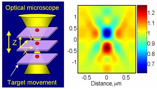 |
| This schematic (left) shows how a TSOM image is acquired. Using an optical microscope, several images of a 60 nanometer gold particle sample (shown in red) are taken at different focal positions and stacked together. The computer-created image on the right shows the resultant TSOM image.
Credit: NIST |
Abstract:
A novel technique* under development at the National Institute of Standards and Technology (NIST) uses a relatively inexpensive optical microscope to quickly and cheaply analyze nanoscale dimensions with nanoscale measurement sensitivity. Termed "Through-focus Scanning Optical Microscope" (TSOM) imaging, the technique has potential applications in nanomanufacturing, semiconductor process control and biotechnology.
Nanoscale Dimensioning Is Fast, Cheap with New NIST Optical Technique
GAITHERSBURG, MD | Posted on October 28th, 2008Optical microscopes are not widely considered for checking nanoscale (below 100 nanometers) dimensions because of the limitation imposed by wavelength of light�you can't get a precise image with a probe three times the object's size. NIST researcher Ravikiran Attota gets around this, paradoxically, by considering lots of "bad" (out-of-focus) images. "This imaging uses a set of blurry, out-of-focus optical images for nanometer dimensional measurement sensitivity," he says. Instead of repeatedly focusing on a sample to acquire one best image, the new technique captures a series of images with an optical microscope at different focal positions and stacks them one on top of the other to create the TSOM image. A computer program Attota developed analyzes the image.
While Attota believes this simple technique can be used in a variety of applications, he has worked with two. The TSOM image can compare two nanoscale objects such as silicon lines on an integrated circuit. The software "subtracts" one image from the other. This enables sensitivity to dimensional differences at the nanoscale�line height, width or side-wall angle. Each type of difference generates a distinct signal.
TSOM has also been theoretically evaluated in another quality control application. Medical researchers are studying the use of gold nanoparticles to deliver advanced pharmaceuticals to specific locations within the human body. Perfect size will be critical. To address this application, a TSOM image of a gold nanoparticle can be taken and compared to a library of simulated images to obtain "best-match" images with the intent of determining if each nanoparticle passes or fails.
This new imaging technology requires a research-quality optical microscope, a camera and a microscope stage that can move at preset distances. "The setup is easily under $50,000, which is much less expensive than electron or probe microscopes currently used for measuring materials at the nanoscale," Attota explains. "This method is another approach to extend the range of optical microscopy from microscale to nanoscale dimensional analysis." So far, sensitivity to a 3 nm difference in line widths has been demonstrated in the laboratory.
* R. Attota, T.A. Germer and R.M. Silver. Through-focus scanning-optical-microscope imaging method for nanoscale dimensional analysis, Optics Letters 33, 1990 (2008).
####
About NIST
Founded in 1901, NIST is a non-regulatory federal agency within the U.S. Department of Commerce. NIST's mission is to promote U.S. innovation and industrial competitiveness by advancing measurement science, standards, and technology in ways that enhance economic security and improve our quality of life.
For more information, please click here
Contacts:
Evelyn Brown
(301) 975-5661
Copyright © NIST
If you have a comment, please Contact us.Issuers of news releases, not 7th Wave, Inc. or Nanotechnology Now, are solely responsible for the accuracy of the content.
Bookmark:


| Related News Press |
News and information
![]() Quantum computer improves AI predictions April 17th, 2026
Quantum computer improves AI predictions April 17th, 2026
![]() Flexible sensor gains sensitivity under pressure April 17th, 2026
Flexible sensor gains sensitivity under pressure April 17th, 2026
![]() A reusable chip for particulate matter sensing April 17th, 2026
A reusable chip for particulate matter sensing April 17th, 2026
![]() Detecting vibrational quantum beating in the predissociation dynamics of SF6 using time-resolved photoelectron spectroscopy April 17th, 2026
Detecting vibrational quantum beating in the predissociation dynamics of SF6 using time-resolved photoelectron spectroscopy April 17th, 2026
Imaging
![]() Simple algorithm paired with standard imaging tool could predict failure in lithium metal batteries August 8th, 2025
Simple algorithm paired with standard imaging tool could predict failure in lithium metal batteries August 8th, 2025
Discoveries
![]() Quantum computer improves AI predictions April 17th, 2026
Quantum computer improves AI predictions April 17th, 2026
![]() Flexible sensor gains sensitivity under pressure April 17th, 2026
Flexible sensor gains sensitivity under pressure April 17th, 2026
![]() A reusable chip for particulate matter sensing April 17th, 2026
A reusable chip for particulate matter sensing April 17th, 2026
![]() Detecting vibrational quantum beating in the predissociation dynamics of SF6 using time-resolved photoelectron spectroscopy April 17th, 2026
Detecting vibrational quantum beating in the predissociation dynamics of SF6 using time-resolved photoelectron spectroscopy April 17th, 2026
Announcements
![]() A fundamentally new therapeutic approach to cystic fibrosis: Nanobody repairs cellular defect April 17th, 2026
A fundamentally new therapeutic approach to cystic fibrosis: Nanobody repairs cellular defect April 17th, 2026
![]() UC Irvine physicists discover method to reverse �quantum scrambling� : The work addresses the problem of information loss in quantum computing system April 17th, 2026
UC Irvine physicists discover method to reverse �quantum scrambling� : The work addresses the problem of information loss in quantum computing system April 17th, 2026
Tools
![]() Metasurfaces smooth light to boost magnetic sensing precision January 30th, 2026
Metasurfaces smooth light to boost magnetic sensing precision January 30th, 2026
![]() From sensors to smart systems: the rise of AI-driven photonic noses January 30th, 2026
From sensors to smart systems: the rise of AI-driven photonic noses January 30th, 2026
![]() Japan launches fully domestically produced quantum computer: Expo visitors to experience quantum computing firsthand August 8th, 2025
Japan launches fully domestically produced quantum computer: Expo visitors to experience quantum computing firsthand August 8th, 2025
|
|
||
|
|
||
| The latest news from around the world, FREE | ||
|
|
||
|
|
||
| Premium Products | ||
|
|
||
|
Only the news you want to read!
Learn More |
||
|
|
||
|
Full-service, expert consulting
Learn More |
||
|
|
||