Home > Press > Lensless camera uses X-rays to view nanoscale materials and biological specimens
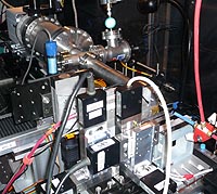 |
| Argonne scientists and collaborators used high energy X-rays from the Advanced Photon Source to create detailed images of nanoscale materials. The scientists are working to develop a dedicated facility for the process at Argonne. |
Abstract:
X-rays have been used for decades to take pictures of broken bones, but scientists at the U.S. Department of Energy's (DOE) Argonne National Laboratory and their collaborators have developed a lensless X-ray technique that can take images of ultra-small structures buried in nanoparticles and nanomaterials, and features within whole biological cells such as cellular nuclei.
Lensless camera uses X-rays to view nanoscale materials and biological specimens
ARGONNE, IL | Posted on February 20th, 2008Argonne scientists along with scientists from the University of California at Los Angeles, the University of Melbourne, La Trobe University and the Australian Synchrotron developed a way to examine internal and buried structures in micrometer-sized samples on the scale of nanometers. This is important to the understanding of how materials behave electrically, magnetically and under thermal and mechanical stress. Application of this capability to biology and biomedicine could contribute to our understanding of disease and its eradication, healing after injury, cancer and cell death.
X-rays are ideally suited for nanoscale imaging because of their ability to penetrate the interior of the object, but their resolution has traditionally been limited by lens technology. The new lensless technique being developed at Argonne avoids this limitation.
"There is no lens involved at all," said Ian McNulty, the lead Argonne author on a new publication on this work appearing in the journal Physical Review Letters. "Instead, a computer uses sophisticated algorithms to reconstruct the image. We expect this technique will enhance our understanding of many problems in materials and biological research." The technique can be extended beyond the current resolution of about 20 nanometers to image the internal structure of micrometer-sized samples at finer resolution, reaching deep into the nanometer scale.
Other types of microscopes, such as electron microscopes, can image structural details on the nanometer scale, but once the sample reaches sizes of a few micrometers and larger, the usefulness of these instruments to probe its internal structure is limited. In many cases, only the surface of the sample can be studied, or the sample must be sliced to view its interior, which can be destructive.
A collaborative team comprising members of the X-ray Microscopy and Imaging Group at Argonne's Advanced Photon Source (APS) and a team led by Professor John Miao at the University of California at Los Angeles developed a powerful new extension of the new lensless imaging technique that enables high resolution imaging of a specific element buried inside a sample.
The key is the high intensity X-ray beams created at the APS at Argonne. An intense, coherent X-ray beam collides with the sample, creating a diffraction pattern which is recorded by a charge coupled device (CCD) camera. The X-ray energy is tuned to an atomic resonance of a target element in the sample. Using sophisticated phase-recovery algorithms, a computer reconstructs an image of the specimen that highlights the presence of the element. The result is an image of the internal architecture of the sample at nanometer resolution and without destructive slicing. By using X-ray energies that coincide with an atomic absorption edge, the imaging process can distinguish between different elements in the sample.
If the nucleus or other parts of a cell are labeled with protein specific tags, it can be imaged within whole cells at a resolution far greater than that of ordinary microscopes.
Another application of this new method of imaging includes the burgeoning field of nanoengineering, which endeavors to develop more efficient catalysts for the petrochemical and energy industries and materials with electrically programmable mechanical, thermal and other properties.
"There are only a handful of places in the world this can be done and APS is the only place in the United States at these X-ray energies," X-ray Microscopy and Imaging Group Leader Qun Shen said. "We would eventually like to create a dedicated, permanent laboratory facility at the APS for this imaging technique that can be used by scientists on a routine basis."
A dedicated facility would require building an additional beamline at the APS, which currently has 34 sectors, each containing one or more beamlines.
This research was funded by the Department of Energy's Office of Basic Energy Sciences as part of its mission to foster and support fundamental research to expand the scientific foundations for new and improved energy technologies, and by the National Science Foundation.
####
About Argonne National Laboratory
Argonne National Laboratory brings the world's brightest scientists and engineers together to find exciting and creative new solutions to pressing national problems in science and technology. The nation's first national laboratory, Argonne conducts leading-edge basic and applied scientific research in virtually every scientific discipline. Argonne researchers work closely with researchers from hundreds of companies, universities, and federal, state and municipal agencies to help them solve their specific problems, advance America 's scientific leadership and prepare the nation for a better future. With employees from more than 60 nations, Argonne is managed by UChicago Argonne, LLC for the U.S. Department of Energy's Office of Science.
For more information, please click here
Contacts:
Brock Cooper
630/252-5565
Argonne
Copyright © Argonne National Laboratory
If you have a comment, please Contact us.Issuers of news releases, not 7th Wave, Inc. or Nanotechnology Now, are solely responsible for the accuracy of the content.
Bookmark:


| Related News Press |
News and information
![]() Quantum computer improves AI predictions April 17th, 2026
Quantum computer improves AI predictions April 17th, 2026
![]() Flexible sensor gains sensitivity under pressure April 17th, 2026
Flexible sensor gains sensitivity under pressure April 17th, 2026
![]() A reusable chip for particulate matter sensing April 17th, 2026
A reusable chip for particulate matter sensing April 17th, 2026
![]() Detecting vibrational quantum beating in the predissociation dynamics of SF6 using time-resolved photoelectron spectroscopy April 17th, 2026
Detecting vibrational quantum beating in the predissociation dynamics of SF6 using time-resolved photoelectron spectroscopy April 17th, 2026
Imaging
![]() Simple algorithm paired with standard imaging tool could predict failure in lithium metal batteries August 8th, 2025
Simple algorithm paired with standard imaging tool could predict failure in lithium metal batteries August 8th, 2025
Discoveries
![]() Quantum computer improves AI predictions April 17th, 2026
Quantum computer improves AI predictions April 17th, 2026
![]() Flexible sensor gains sensitivity under pressure April 17th, 2026
Flexible sensor gains sensitivity under pressure April 17th, 2026
![]() A reusable chip for particulate matter sensing April 17th, 2026
A reusable chip for particulate matter sensing April 17th, 2026
![]() Detecting vibrational quantum beating in the predissociation dynamics of SF6 using time-resolved photoelectron spectroscopy April 17th, 2026
Detecting vibrational quantum beating in the predissociation dynamics of SF6 using time-resolved photoelectron spectroscopy April 17th, 2026
Announcements
![]() A fundamentally new therapeutic approach to cystic fibrosis: Nanobody repairs cellular defect April 17th, 2026
A fundamentally new therapeutic approach to cystic fibrosis: Nanobody repairs cellular defect April 17th, 2026
![]() UC Irvine physicists discover method to reverse �quantum scrambling� : The work addresses the problem of information loss in quantum computing system April 17th, 2026
UC Irvine physicists discover method to reverse �quantum scrambling� : The work addresses the problem of information loss in quantum computing system April 17th, 2026
Tools
![]() Metasurfaces smooth light to boost magnetic sensing precision January 30th, 2026
Metasurfaces smooth light to boost magnetic sensing precision January 30th, 2026
![]() From sensors to smart systems: the rise of AI-driven photonic noses January 30th, 2026
From sensors to smart systems: the rise of AI-driven photonic noses January 30th, 2026
![]() Japan launches fully domestically produced quantum computer: Expo visitors to experience quantum computing firsthand August 8th, 2025
Japan launches fully domestically produced quantum computer: Expo visitors to experience quantum computing firsthand August 8th, 2025
|
|
||
|
|
||
| The latest news from around the world, FREE | ||
|
|
||
|
|
||
| Premium Products | ||
|
|
||
|
Only the news you want to read!
Learn More |
||
|
|
||
|
Full-service, expert consulting
Learn More |
||
|
|
||