Home > Press > Higher-resolution imaging of living, moving cells using plasmonic metasurfaces: Metasurface of self-assembled gold nanoparticles shown to improve resolution of fluorescence images of living cells under a widefield optical microscope to the theoretical limit
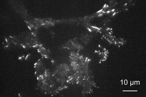 |
| A mouse fibroblast cell imaged on a metasurface made of gold nanoparticles under a total internal reflection fluorescence (TIRF) microscope exhibits enhanced and confined emission from light-emitting paxillin protein at focal adhesions near the membrane. Researchers at Kyushu University have shown that such metasurfaces can be used with conventional fluorescence microscopes as a simple route to improve the resolution to near the diffraction limit. The use of TIRF conditions helps to reduce stray emission from deep within the cell to further improve contrast of structures near the metasurface. CREDIT Kaoru Tamada, Kyushu University |
Abstract:
In the quest to image exceedingly small structures and phenomenon with higher precision, scientists have been pushing the limits of optical microscope resolution, but these advances often come with increased complication and cost.
Higher-resolution imaging of living, moving cells using plasmonic metasurfaces: Metasurface of self-assembled gold nanoparticles shown to improve resolution of fluorescence images of living cells under a widefield optical microscope to the theoretical limit
Fukuoka, Japan | Posted on November 6th, 2020Now, researchers in Japan have shown that a glass surface embedded with self-assembled gold nanoparticles can improve resolution with little added cost even using a conventional widefield microscope, facilitating high-resolution fluorescence microscopy capable of high-speed imaging of living cells.
Because optical microscopes magnify light to obtain detailed images of a structure, the size of objects that can be distinguished has long been limited by diffraction--a property of light that causes it to spread when passing through an opening.
Researchers have been developing techniques to overcome these limits with highly advanced optical systems, but many of them depend on the use of strong lasers, which can damage or even kill living cells, and scanning of the sample or processing of multiple images, which inhibits real-time imaging.
"Recent techniques can produce stunning images, but many of them require highly specialized equipment and are incapable of observing the movement of living cells," says Kaoru Tamada, distinguished professor at Kyushu University's Institute for Materials Chemistry and Engineering.
Imaging cells using real-time fluorescence microscopy methods, Tamada and her group found that they could improve resolution under a conventional widefield microscope to near the diffraction limit just by changing the surface under the cells.
In fluorescence microscopy, cell structures of interest are tagged with molecules that absorb energy from incoming light and, through the process of fluorescence, re-emit it as light of a different color, which is collected to form the image.
Though cells are usually imaged on plain glass, Tamada's group coated the glass surface with a self-assembled layer of gold nanoparticles covered with a thin layer of silicon dioxide, creating a so-called metasurface with special optical properties.
Only 12 nm in diameter, the organized metal nanoparticles exhibit a phenomenon known as localized surface plasmon resonance, which allows the metasurface to collect energy from nearby light-emitting molecules for highly efficient re-emission, thereby producing enhanced emission confined to the 10-nm thick nanoparticle surface.
"By introducing the nanoparticles, we have effectively created a light-emitting plane that is only several nanometers thick," explains Tamada. "Because the light of interest is emitted from such a thin layer, we can better focus on it."
Additional benefits arise from energy transfer to the metasurface being fast, further localizing emission points by reducing diffusion, and the metasurface's high refractive index, which helps to improve resolution according to Abbe's diffraction limit.
Using the metasurface, the researchers imaged in real-time mouse cells known as 3T3 fibroblasts that were genetically engineered to produce a protein called paxillin that is modified to emit green light when excited. Paxillin plays a key role in creating focal adhesions--points where molecules in the cell membrane interact with the outside world.
Illuminating the entire sample with laser light perpendicular to the surface, the researchers were able to image changes in paxillin near the cell membrane with a higher resolution using the metasurface instead of glass.
Tilting the illumination light to achieve total internal reflection, the researchers could obtain images with even higher contrast because most of the illumination light is reflected off the surface with only a small amount reaching the cell side, thereby reducing stray emission produced by illumination penetrating deep into the cell.
Analysis of images recorded every 500 milliseconds with a super-resolution digital camera revealed clear differences in intensity over spots covering only a few pixels, indicating the resolution was about 200 nm--close to the diffraction limit.
Cells could also be imaged longer on the metasurface because the emission was enhanced despite a lower input energy, thereby reducing cell damage over time.
"Metasurfaces are a promising option for improving resolution for researchers around the world using conventional optical microscopes that they already have," comments Tamada.
In addition to continuing to improve the surfaces for use with conventional microscopes, the researchers are also exploring the advantages they can have for more sophisticated microscope systems.
####
For more information, please click here
Contacts:
William J. Potscavage Jr.
81-928-022-138
@KyushuUniv_EN
Copyright © Kyushu University
If you have a comment, please Contact us.Issuers of news releases, not 7th Wave, Inc. or Nanotechnology Now, are solely responsible for the accuracy of the content.
Bookmark:


| Related Links |
| Related News Press |
News and information
![]() Decoding hydrogen‑bond network of electrolyte for cryogenic durable aqueous zinc‑ion batteries January 30th, 2026
Decoding hydrogen‑bond network of electrolyte for cryogenic durable aqueous zinc‑ion batteries January 30th, 2026
![]() COF scaffold membrane with gate‑lane nanostructure for efficient Li+/Mg2+ separation January 30th, 2026
COF scaffold membrane with gate‑lane nanostructure for efficient Li+/Mg2+ separation January 30th, 2026
![]() MXene nanomaterials enter a new dimension Multilayer nanomaterial: MXene flakes created at Drexel University show new promise as 1D scrolls January 30th, 2026
MXene nanomaterials enter a new dimension Multilayer nanomaterial: MXene flakes created at Drexel University show new promise as 1D scrolls January 30th, 2026
Imaging
![]() ICFO researchers overcome long-standing bottleneck in single photon detection with twisted 2D materials August 8th, 2025
ICFO researchers overcome long-standing bottleneck in single photon detection with twisted 2D materials August 8th, 2025
Possible Futures
![]() Decoding hydrogen‑bond network of electrolyte for cryogenic durable aqueous zinc‑ion batteries January 30th, 2026
Decoding hydrogen‑bond network of electrolyte for cryogenic durable aqueous zinc‑ion batteries January 30th, 2026
![]() COF scaffold membrane with gate‑lane nanostructure for efficient Li+/Mg2+ separation January 30th, 2026
COF scaffold membrane with gate‑lane nanostructure for efficient Li+/Mg2+ separation January 30th, 2026
Discoveries
![]() From sensors to smart systems: the rise of AI-driven photonic noses January 30th, 2026
From sensors to smart systems: the rise of AI-driven photonic noses January 30th, 2026
![]() Decoding hydrogen‑bond network of electrolyte for cryogenic durable aqueous zinc‑ion batteries January 30th, 2026
Decoding hydrogen‑bond network of electrolyte for cryogenic durable aqueous zinc‑ion batteries January 30th, 2026
![]() COF scaffold membrane with gate‑lane nanostructure for efficient Li+/Mg2+ separation January 30th, 2026
COF scaffold membrane with gate‑lane nanostructure for efficient Li+/Mg2+ separation January 30th, 2026
Announcements
![]() Decoding hydrogen‑bond network of electrolyte for cryogenic durable aqueous zinc‑ion batteries January 30th, 2026
Decoding hydrogen‑bond network of electrolyte for cryogenic durable aqueous zinc‑ion batteries January 30th, 2026
![]() COF scaffold membrane with gate‑lane nanostructure for efficient Li+/Mg2+ separation January 30th, 2026
COF scaffold membrane with gate‑lane nanostructure for efficient Li+/Mg2+ separation January 30th, 2026
Interviews/Book Reviews/Essays/Reports/Podcasts/Journals/White papers/Posters
![]() Metasurfaces smooth light to boost magnetic sensing precision January 30th, 2026
Metasurfaces smooth light to boost magnetic sensing precision January 30th, 2026
![]() COF scaffold membrane with gate‑lane nanostructure for efficient Li+/Mg2+ separation January 30th, 2026
COF scaffold membrane with gate‑lane nanostructure for efficient Li+/Mg2+ separation January 30th, 2026
Tools
![]() Metasurfaces smooth light to boost magnetic sensing precision January 30th, 2026
Metasurfaces smooth light to boost magnetic sensing precision January 30th, 2026
![]() From sensors to smart systems: the rise of AI-driven photonic noses January 30th, 2026
From sensors to smart systems: the rise of AI-driven photonic noses January 30th, 2026
![]() Japan launches fully domestically produced quantum computer: Expo visitors to experience quantum computing firsthand August 8th, 2025
Japan launches fully domestically produced quantum computer: Expo visitors to experience quantum computing firsthand August 8th, 2025
|
|
||
|
|
||
| The latest news from around the world, FREE | ||
|
|
||
|
|
||
| Premium Products | ||
|
|
||
|
Only the news you want to read!
Learn More |
||
|
|
||
|
Full-service, expert consulting
Learn More |
||
|
|
||