Home > Press > Tuberculosis bacterium is double-protected
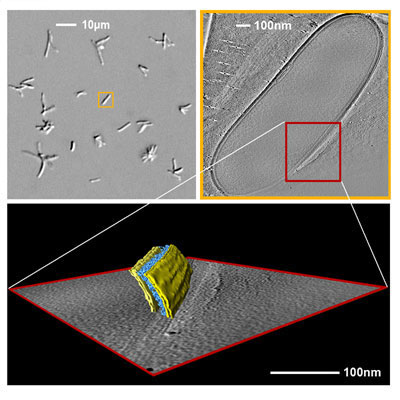 |
| Light and electron tomographic images of Mycobacterium bovis BCG. Top left: Several mycobacteria in the light microscope with about 1000 times magnification. Top right: Longitudinal section through a three-dimensionally reconstructed bacterial cell of 1.5 �m in length. The data were recorded in an electron microscope using the technique of cryo-electron tomography. The white bar indicates 100 nanometer or 0.1 micron. Bottom: 3-D structure of a section through the mycobacterial cell envelope. The structural details are color-coded. In yellow: the inner (left) and the outer (right) lipid bilayer. In blue: cell wall polymers that bind the mycolic acids. The cell envelope is 35 nanometers thick.
Image: Christian Hoffmann/Harald Engelhardt, MPI of Biochemistry, Martinsried, Germany |
Abstract:
The first 3-D images that disclosure a double membrane surrounding mycobacteria were recorded by Martinsried scientists, ending a long scientific debate about the mycobacterial outer membrane and opening new pathways to improve the development of chemotherapeutic substances against tuberculosis (PNAS March 2008).
Tuberculosis bacterium is double-protected
Munich, Germany | Posted on March 7th, 2008 Robert Koch who detected the tuberculosis causative agent in March 1882 described the contemporary situation: "Statistics tells us that one in seven of all humans dies of tuberculosis�" Even today, ten million people suffer from the disease yearly, and every day, Mycobacterium tuberculosis causes the death of about 4000 patients. Medical treatment is lengthy and protection through vaccinations is, today as before, insufficient. This is why research groups worldwide study the �acid-fast rods', which are protected by a complex and hardly penetrable cell wall. Its special structure is the reason for the resistance to external factors and for the inefficient uptake of antibacterial substances.
It has been known for some time that long-chained, strongly bound fatty acids - the mycolic acids - are necessary to preserve the resistance properties of the cell wall. But even 125 years after Koch's discovery, our knowledge of the mycobacterial cell envelope is incomplete and characterized by contradictory hypotheses. Until recently, scientists assumed that mycolic acids form a closed layer, or that they comprise the inner part of a considerably thick and asymmetrical membrane. Now, Harald Engelhardt and his group at the Max Planck Institute of Biochemistry in Martinsried have proved that the outer cell wall layer consists of a distinct lipid bilayer. Its structure, however, is hardly compatible with the current view of the cell wall architecture.
Christian Hoffman, a PhD student in Harald Engelhardt's lab, investigated the cell structure of Mycobacterium smegmatis and Mycobacterium bovis BCG, a close relative to the tuberculosis bacterium, in the electron microscope. The scientists were able to obtain 3-D images of the bilayer structure from intact cells by means of cryo-electron tomography, a technique that was developed at the institute in Martinsried. The method requires projection data from different angles of a shock-frozen cell (-190 �C) that are optimized for the number, contrast and focus of the images. In order to avoid radiation damage, the cell must only be exposed to the electron beam for a limited period of time. The images are thus noisy and lack contrast. The Department for Molecular Structural Biology, headed by Professor Wolfgang Baumeister, and in this case especially J�rgen Plitzko, pioneers development and research of cryo-electron tomography, which is a valuable technique to investigate structures of intact cells in a close-to-life state.
Hoffmann and his colleagues observed a more symmetrical and significantly thinner mycobacterial membrane than previously expected. The researchers therefore probed their results by electron microscopy of ultrathin cryosections of frozen cells (each section 35 millionth of a millimeter in thickness), which had not been treated further, and could confirm their findings. The researchers can now satisfactorily explain how the pore proteins are embedded in outer membrane of Mycobacterium smegmatis. The molecular structure of these proteins did not fit to the existing models of the mycobacterial cell wall.
Harald Engelhardt, the leader of the research project, agrees with previous hypotheses insofar as mycolic acids anchor the outer membrane to the cell wall. "But the membrane is probably not structured the way we thought. The mycolic and other fatty acids must be organized differently in the cell membrane than previously assumed." The Martinsried microbiologists and structural researchers now see the need for a more detailed study of the mycobacterial outer membrane. The recent findings provide an appropriate basis for such inquiries. Because now, distinct studies investigating the translocation of molecular substances across the outer membrane have been made possible, which should also be useful for the development of chemotherapeutic drugs. Engelhardt: "After all, the drugs must pass through cell wall as effectively as possible, and a better understanding of the mycobacterial cell envelope will certainly be helpful."
####
About Max Planck Society
The research institutes of the Max Planck Society perform basic research in the interest of the general public in the natural sciences, life sciences, social sciences, and the humanities. In particular, the Max Planck Society takes up new and innovative research areas that German universities are not in a position to accommodate or deal with adequately. These interdisciplinary research areas often do not fit into the university organization, or they require more funds for personnel and equipment than those available at universities. The variety of topics in the natural sciences and the humanities at Max Planck Institutes complement the work done at universities and other research facilities in important research fields. In certain areas, the institutes occupy key positions, while other institutes complement ongoing research. Moreover, some institutes perform service functions for research performed at universities by providing equipment and facilities to a wide range of scientists, such as telescopes, large-scale equipment, specialized libraries, and documentary resources.
For more information, please click here
Contacts:
Max Planck Society
for the Advancement of Science
Press and Public Relations Department
Hofgartenstrasse 8
D-80539 Munich
Germany
PO Box 10 10 62
D-80084 Munich
Phone: +49-89-2108-1276
Fax: +49-89-2108-1207
E-mail:
Internet: www.mpg.de/english/
Responsibility for content:
Dr. Bernd Wirsing (-1276)
Executive Editor:
Barbara Abrell (-1416)
Copyright © Max Planck Society
If you have a comment, please Contact us.Issuers of news releases, not 7th Wave, Inc. or Nanotechnology Now, are solely responsible for the accuracy of the content.
Bookmark:


| Related News Press |
News and information
![]() Quantum computer improves AI predictions April 17th, 2026
Quantum computer improves AI predictions April 17th, 2026
![]() Flexible sensor gains sensitivity under pressure April 17th, 2026
Flexible sensor gains sensitivity under pressure April 17th, 2026
![]() A reusable chip for particulate matter sensing April 17th, 2026
A reusable chip for particulate matter sensing April 17th, 2026
![]() Detecting vibrational quantum beating in the predissociation dynamics of SF6 using time-resolved photoelectron spectroscopy April 17th, 2026
Detecting vibrational quantum beating in the predissociation dynamics of SF6 using time-resolved photoelectron spectroscopy April 17th, 2026
Imaging
![]() Simple algorithm paired with standard imaging tool could predict failure in lithium metal batteries August 8th, 2025
Simple algorithm paired with standard imaging tool could predict failure in lithium metal batteries August 8th, 2025
Nanomedicine
![]() A fundamentally new therapeutic approach to cystic fibrosis: Nanobody repairs cellular defect April 17th, 2026
A fundamentally new therapeutic approach to cystic fibrosis: Nanobody repairs cellular defect April 17th, 2026
![]() New molecular technology targets tumors and simultaneously silences two �undruggable� cancer genes August 8th, 2025
New molecular technology targets tumors and simultaneously silences two �undruggable� cancer genes August 8th, 2025
![]() New imaging approach transforms study of bacterial biofilms August 8th, 2025
New imaging approach transforms study of bacterial biofilms August 8th, 2025
![]() Electrifying results shed light on graphene foam as a potential material for lab grown cartilage June 6th, 2025
Electrifying results shed light on graphene foam as a potential material for lab grown cartilage June 6th, 2025
Discoveries
![]() Quantum computer improves AI predictions April 17th, 2026
Quantum computer improves AI predictions April 17th, 2026
![]() Flexible sensor gains sensitivity under pressure April 17th, 2026
Flexible sensor gains sensitivity under pressure April 17th, 2026
![]() A reusable chip for particulate matter sensing April 17th, 2026
A reusable chip for particulate matter sensing April 17th, 2026
![]() Detecting vibrational quantum beating in the predissociation dynamics of SF6 using time-resolved photoelectron spectroscopy April 17th, 2026
Detecting vibrational quantum beating in the predissociation dynamics of SF6 using time-resolved photoelectron spectroscopy April 17th, 2026
Announcements
![]() A fundamentally new therapeutic approach to cystic fibrosis: Nanobody repairs cellular defect April 17th, 2026
A fundamentally new therapeutic approach to cystic fibrosis: Nanobody repairs cellular defect April 17th, 2026
![]() UC Irvine physicists discover method to reverse �quantum scrambling� : The work addresses the problem of information loss in quantum computing system April 17th, 2026
UC Irvine physicists discover method to reverse �quantum scrambling� : The work addresses the problem of information loss in quantum computing system April 17th, 2026
|
|
||
|
|
||
| The latest news from around the world, FREE | ||
|
|
||
|
|
||
| Premium Products | ||
|
|
||
|
Only the news you want to read!
Learn More |
||
|
|
||
|
Full-service, expert consulting
Learn More |
||
|
|
||