Home > Nanotechnology Columns > Cristian Orfescu > The Beginnings of NanoArt
 |
Cris Orfescu Founder NanoArt21 |
Abstract:
There are numerous sources that mention the art - nanotechnology intersections from early times of the human history. When did NanoArt come into the picture?
June 6th, 2012
The Beginnings of NanoArt
Art - nanotechnology intersections are mentioned in numerous sources from early times of the human history. When did the NanoArt started? I would say that once the electron microscope became commercially available (late 1930s) the first NanoArt works were created. Apparently, the scientists who were imaging those small structures did not have artistic intentions, but they created images that could be considered artworks and some of them were quite aesthetic.
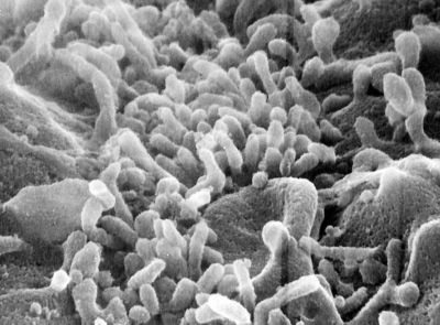 |
| Jamieson, James D. - Single guinea pig pancreatic acinar cell scanning EM apex |
One of the first nanoartists in the history, probably without his intention to create art was George Emil Palade (1912 - 2008), a Romanian cell biologist. Described as "the most influential cell biologist ever" (Hopkins, 2008), he was awarded in 1974 the Nobel Prize in Physiology and Medicine together with Albert Claude and Christian de Duve for innovations in electron microscopy and discoveries concerning the structural and functional organization of the cell that laid the foundations of modern molecular cell biology. The most notable discovery was the ribosomes of the endoplasmic reticulum
described by Palade first in 1955.
 |
| Ohad, I. - Unicellular Organisms Yellow Chlamydomonas |
The George E. Palade Electron Microscopy Slide Collection of electron microscopy images at Harvey Cushing/John Hay Whitney Medical Library, Yale University, derived from high-resolution images scanned by James D. Jamieson and is freely available to students and scientists worldwide.
 |
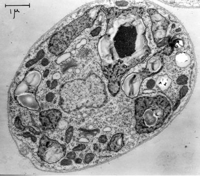
| Palade, George E. - Mitochondria Guniea Pig Pancreas lipid droplet |
The collection of images includes some of the earliest electron micrographs taken by George Palade and his collaborators at the Rockefeller University (1945-1973) and at Yale University (1973-1990). Electron micrographs taken by Marilyn Farquhar (the glomerular basement membrane in renal filtration), Maya and Nicolae Simeonescu (capillary endothelium), James Jamieson (secretory pathway in the exocrine pancreas; atrial granules), Lucien Caro (electron microscopic autoradiography), Philip Siekevitz, John Bergeron and Japoco Meldolesi (microsomes, Golgi fractions), and Sanford Palay (synapses) are part of the collection.
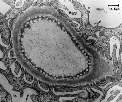 |
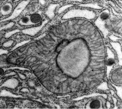
| Farquhar, Marilyn G. - Kidney Rat glomerular filtration barrier tangential section fenestrated endothelium |
All these scientists, consciously or not, created valuable scientific imagery, and in the same time they pioneered in a new discipline, NanoArt.
Bookmark:


|
|
||
|
|
||
| The latest news from around the world, FREE | ||
|
|
||
|
|
||
| Premium Products | ||
|
|
||
|
Only the news you want to read!
Learn More |
||
|
|
||
|
Full-service, expert consulting
Learn More |
||
|
|
||