Home > Press > High resolution without particle accelerator: A first for physics -- University of Jena physicists are first to achieve optical coherence tomography with XUV radiation at laboratory scale
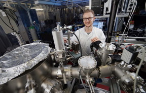 |
| Silvio Fuchs in a laboratory of the Institute of Optics and Quantum Electronics of the Friedrich Schiller University Jena. CREDIT Photo: Jan-Peter Kasper/FSU Jena |
Abstract:
A visit to the optometrist often involves optical coherence tomography. This imaging process uses infrared radiation to penetrate the layers of the retina and examine it more closely in three dimensions, without having to touch the eye at all. This allows eye specialists to diagnose diseases such as glaucoma without any physical intervention. However, this method would have even greater potential for science if a shorter radiation wavelength were used, thus allowing a higher resolution of the image. Physicists at Friedrich Schiller University Jena (Germany) have now achieved just that and they have reported their research findings in the latest issue of the specialist journal "Optica" (DOI: 10.1364/OPTICA.4.000903).
High resolution without particle accelerator: A first for physics -- University of Jena physicists are first to achieve optical coherence tomography with XUV radiation at laboratory scale
Jena, Germany | Posted on August 7th, 2017First XUV coherence tomography at laboratory scale
For the first time, the University physicists used extreme ultraviolet radiation (XUV) for this process, which was generated in their own laboratory, and they were thus able to perform the first XUV coherence tomography at laboratory scale. This radiation has a wavelength of between 20 and 40 nanometres -- from which it is therefore just a small step to the X-ray range. "Large-scale equipment, that is to say particle accelerators such as the German Elektronen-Synchotron in Hamburg, are usually necessary for generating XUV radiation," says Silvio Fuchs of the Institute of Optics and Quantum Electronics of the Jena University. "This makes such a research method very complex and costly, and only available to a few researchers." The physicists from Jena have already demonstrated this method at large research facilities, but they have now found a possibility for applying it at a smaller scale. In this approach, they focus an ultrashort, very intense infrared laser in a noble gas, for example argon or neon. "The electrons in the gas are accelerated by means of an ionisation process," explains Fuchs. "They then emit the XUV radiation." It is true that this method is very inefficient, as only a millionth part of the laser radiation is actually transformed from infrared into the extreme ultraviolet range, but this loss can be offset by the use of very powerful laser sources. "It's a simple calculation: the more we put in, the more we get out," adds Fuchs.
Strong image contrasts are produced
The advantage of XUV coherence tomography is that, in addition to the very high resolution, the radiation interacts strongly with the sample, because differrent substances react differently to light. Some absorb more light and others less. This produces strong contrasts in the images, which provide the researchers with important information, for example regarding the material composition of the object being examined. "For example, we have created three-dimensional images of silicon chips, in a non-destructive way, on which we can distinguish the substrate clearly from structures consisting of other materials," adds Silvio Fuchs. "If this procedure were applied in biology -- for investigating cells, for example, which is one of our aims -- it would not be necessary to colour samples, as is normal practice in other high-resolution microscopy methods. Elements such as carbon, oxygen and nitrogen would themselves provide the contrast." Before that is possible, however, the physicists of the University of Jena still have some work to do. "With the light sources we have at the moment, we can achieve a depth resolution down to 24 nanometres. Although this is sufficient for producing images of small structures, for example in semiconductors, the structure sizes of current chips are in some cases already smaller. However, with new, even more powerful lasers, it should be possible in future to achieve a depth resolution of as little as three nanometres with this method," notes Fuchs. "We have shown in principle that it is possible to use this method at laboratory scale."
The long-term aim could ultimately be to develop a cost-effective and user-friendly device combining the laser with the microscope, which would enable the semiconductor industry or biological laboratories to use this imaging technique with ease.
####
For more information, please click here
Contacts:
Axel Burchardt
49-364-193-1031
Silvio Fuchs
Institute of Optics and Quantum Electronics
Friedrich Schiller University Jena
Max-Wien-Platz 1, 07743 Jena, Germany
Phone: +49-0-3641 / 947615
Copyright © Friedrich-Schiller-Universit�t Jena
If you have a comment, please Contact us.Issuers of news releases, not 7th Wave, Inc. or Nanotechnology Now, are solely responsible for the accuracy of the content.
Bookmark:


| Related Links |
| Related News Press |
News and information
![]() Simulating magnetization in a Heisenberg quantum spin chain April 5th, 2024
Simulating magnetization in a Heisenberg quantum spin chain April 5th, 2024
![]() NRL charters Navy�s quantum inertial navigation path to reduce drift April 5th, 2024
NRL charters Navy�s quantum inertial navigation path to reduce drift April 5th, 2024
![]() Discovery points path to flash-like memory for storing qubits: Rice find could hasten development of nonvolatile quantum memory April 5th, 2024
Discovery points path to flash-like memory for storing qubits: Rice find could hasten development of nonvolatile quantum memory April 5th, 2024
Imaging
![]() Nanoscale CL thermometry with lanthanide-doped heavy-metal oxide in TEM March 8th, 2024
Nanoscale CL thermometry with lanthanide-doped heavy-metal oxide in TEM March 8th, 2024
![]() The USTC realizes In situ electron paramagnetic resonance spectroscopy using single nanodiamond sensors November 3rd, 2023
The USTC realizes In situ electron paramagnetic resonance spectroscopy using single nanodiamond sensors November 3rd, 2023
![]() Observation of left and right at nanoscale with optical force October 6th, 2023
Observation of left and right at nanoscale with optical force October 6th, 2023
Possible Futures
![]() Discovery points path to flash-like memory for storing qubits: Rice find could hasten development of nonvolatile quantum memory April 5th, 2024
Discovery points path to flash-like memory for storing qubits: Rice find could hasten development of nonvolatile quantum memory April 5th, 2024
![]() With VECSELs towards the quantum internet Fraunhofer: IAF achieves record output power with VECSEL for quantum frequency converters April 5th, 2024
With VECSELs towards the quantum internet Fraunhofer: IAF achieves record output power with VECSEL for quantum frequency converters April 5th, 2024
Discoveries
![]() Chemical reactions can scramble quantum information as well as black holes April 5th, 2024
Chemical reactions can scramble quantum information as well as black holes April 5th, 2024
![]() New micromaterial releases nanoparticles that selectively destroy cancer cells April 5th, 2024
New micromaterial releases nanoparticles that selectively destroy cancer cells April 5th, 2024
![]() Utilizing palladium for addressing contact issues of buried oxide thin film transistors April 5th, 2024
Utilizing palladium for addressing contact issues of buried oxide thin film transistors April 5th, 2024
Announcements
![]() NRL charters Navy�s quantum inertial navigation path to reduce drift April 5th, 2024
NRL charters Navy�s quantum inertial navigation path to reduce drift April 5th, 2024
![]() Discovery points path to flash-like memory for storing qubits: Rice find could hasten development of nonvolatile quantum memory April 5th, 2024
Discovery points path to flash-like memory for storing qubits: Rice find could hasten development of nonvolatile quantum memory April 5th, 2024
Interviews/Book Reviews/Essays/Reports/Podcasts/Journals/White papers/Posters
![]() Simulating magnetization in a Heisenberg quantum spin chain April 5th, 2024
Simulating magnetization in a Heisenberg quantum spin chain April 5th, 2024
![]() Discovery points path to flash-like memory for storing qubits: Rice find could hasten development of nonvolatile quantum memory April 5th, 2024
Discovery points path to flash-like memory for storing qubits: Rice find could hasten development of nonvolatile quantum memory April 5th, 2024
Tools
![]() Ferroelectrically modulate the Fermi level of graphene oxide to enhance SERS response November 3rd, 2023
Ferroelectrically modulate the Fermi level of graphene oxide to enhance SERS response November 3rd, 2023
![]() The USTC realizes In situ electron paramagnetic resonance spectroscopy using single nanodiamond sensors November 3rd, 2023
The USTC realizes In situ electron paramagnetic resonance spectroscopy using single nanodiamond sensors November 3rd, 2023
Photonics/Optics/Lasers
![]() With VECSELs towards the quantum internet Fraunhofer: IAF achieves record output power with VECSEL for quantum frequency converters April 5th, 2024
With VECSELs towards the quantum internet Fraunhofer: IAF achieves record output power with VECSEL for quantum frequency converters April 5th, 2024
![]() Nanoscale CL thermometry with lanthanide-doped heavy-metal oxide in TEM March 8th, 2024
Nanoscale CL thermometry with lanthanide-doped heavy-metal oxide in TEM March 8th, 2024
![]() Optically trapped quantum droplets of light can bind together to form macroscopic complexes March 8th, 2024
Optically trapped quantum droplets of light can bind together to form macroscopic complexes March 8th, 2024
![]() HKUST researchers develop new integration technique for efficient coupling of III-V and silicon February 16th, 2024
HKUST researchers develop new integration technique for efficient coupling of III-V and silicon February 16th, 2024
|
|
||
|
|
||
| The latest news from around the world, FREE | ||
|
|
||
|
|
||
| Premium Products | ||
|
|
||
|
Only the news you want to read!
Learn More |
||
|
|
||
|
Full-service, expert consulting
Learn More |
||
|
|
||