Home > Press > Wellcome Image Awards 2008
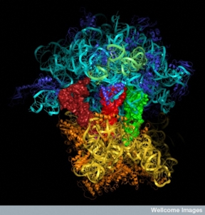 |
| Molecular model of a ribosome Credit:MRC Lab of Molecular Biology |
Abstract:
Bold, beautiful and groundbreaking, this year's Wellcome Image Awards delve deep into our understanding of modern medicine and science.
Wellcome Image Awards 2008
London, UK | Posted on March 12th, 2008Dazzling and brightly coloured images depict subject matters as diverse as breast cancer cells, Clostridium difficile and ruptured blood vessels, and exemplify recent advances in science and medicine.
Each year, the Wellcome Trust runs the Wellcome Image Awards ceremony (previously the Biomedical Image Awards) to recognise the scientists who have created stunning and beautiful images as part of their own research and made them available for public use through the Wellcome Library's image repository, Wellcome Images.
The award-winning images represent a fraction of the images contained in Wellcome Images - most are taken from the Wellcome Library's vast collection of resources on the history and culture of medicine.
Vivienne Parry will present the Awards at a special ceremony at Wellcome Collection on Tuesday 11 March at 18.30 and the 22 winning images will be on display in the Wellcome Collection Atrium from 12 March onwards.
Wellcome Image Awards 2008 ceremony: Tuesday 11 March 2008, 18.30-20.30
Wellcome Image Awards: 12 March 2008 - Summer 2008
Venue: Wellcome Collection, 183 Euston Road, London NW1 2BE Admission free
Opening times: Mon.-Wed., Fri.-Sat.: 10.00-18.00. Thurs.: 10.00-22.00. Sun.: 11.00-18.00 (NB exhibitions galleries are closed on Monday)
Each of the winning images has been carefully selected by a panel of judges including Vivienne Parry (science broadcaster), Beau Lotto (neuroscientist and expert in visual perception), Dr Alice Roberts (medical doctor and presenter of the BBC's 'Don't Die Young') and Rachel Dickens (Deputy Art Editor of 'BBC Focus' magazine).
Vivienne Parry explains: "It was very hard deciding on the winning images, not only because all of them are stunning but also because we had to consider their scientific content, the skills needed to make them and also how good they were at making complex science accessible."
Catherine Draycott, Head of Wellcome Images, explains: "We are delighted with the results of this year's awards. The winning scientists have created stunning and beautiful images as part of their own research which can themselves be used widely in communicating science to all."
The 22 award-winning images for this year all have a fascinating story to tell, including:
Red blood cells oozing from a ruptured vessel - revealing how a genetic mutation can lead to haemorrhaging similar to that seen in the blood vessels that feed developing cancers, by Anne Weston, Cancer Research UK.
The image of a circle of DNA, created using a molecular dynamics simulation to study whether clay nanomaterials could have played a role in the origins of life by protecting DNA in extreme conditions, has been made by Mary-Ann Thyveetil of University College London.
A mouse embryo, using a new technique - optical projection tomography - to examine internal structures, without the need for cutting sections, by James Sharpe Human Genetics Unit in Edinburgh.
Image of crystals of oxidised vitamin C by Spike Walker reveals the beautiful, almost marine-like shapes created by the crystallisation of this important vitamin. The ease with which vitamin C is oxidised is vitally important in protecting cells from damaging free radicals.
Contact
Mike Findlay
Wellcome Collection Media Officer
T 020 7611 8612
E
Notes to editors
1) Dates and venues
The Wellcome Image Awards 2008 is on show at Wellcome Collection, at 183 Euston Road, London, from 12 March 2008.
It will also be on show in Tokyo at Miraikan, the National Museum of Emerging Science and Innovation. Dates and details to be confirmed.
2) Press images for Wellcome Image Awards 2008
View the award-winning images.
These images are provided for use with stories on Wellcome Image Awards 2008 only. Please ensure that you use the appropriate credit line. Images must not be archived or re-used for any other purpose without the prior written permission of Wellcome Images.
3) Wellcome Images
Wellcome Images allows unlimited access to a vast catalogue of medical images, manuscripts, and illustrations exploring the meaning of medicine, its history and current practice.
All content has been made available under a Creative Commons Licence, which allows users to copy, distribute and display the image, provided the source is fully attributed and it is used for non-commercial purposes.
The images are also available for all media uses; contact Wellcome Images for more information.
Everything from an oil painting of Florence Nightingale and a picture depicting Charles Darwin as an ape, to a photograph of Alexander Fleming in his laboratory are part of this unique collection. The images aid teachers and researchers to illustrate themes from medical and social history through to contemporary healthcare and biomedical science, and to bring complex biomedical concepts to life.
4) The Wellcome Library
The Wellcome Library is an unrivalled resource for the study of the history of medicine, to collection includes: 700 000 books; a film and audio collection of 2500 titles; 600 archival collections; and more than 100 000 paintings, prints and photographs. Over 100 000 images in Wellcome Images have been sourced from the Library's collections. The Library is part of Wellcome Collection.
The Wellcome Trust is the largest charity in the UK. It funds innovative biomedical research, in the UK and internationally, spending over �650 million each year to support the brightest scientists with the best ideas. The Trust supports public debate about biomedical research and its impact on health and wellbeing.
Our former headquarters, the Wellcome Building on London's Euston Road, has been redesigned by Hopkins Architects to become a new �30 million public venue. Free to all, Wellcome Collection explores the connections between medicine, life and art in the past, present and future. The building comprises three galleries, a public events space, the Wellcome Library, a caf�, a bookshop, conference facilities and a members' club.
5) The judging panel
Rachel Dickens has worked as a designer for BBC magazines since 2006 and is the Deputy Art Editor of science and technology magazine 'BBC Focus'. She has an honours degree in illustration and a background in advertising design. In 2007 she was nominated for the PTC New Magazine Designer of the Year Award.
Catherine Draycott has been Head of Wellcome Images, part of the Wellcome Library, since 1992. She is responsible for the development of the department to provide access to a comprehensive collection of images of medicine and its history for both individual users and the media. She has been a judge of the Wellcome Image Awards since their inception in 1997. She is a Director of the British Association of Picture Libraries and Agencies and was its Chairman from 2000 to 2007.
Beau Lotto is a reader in neuroscience and head of lottolab at University College London. His work attempts to understand why the brain 'sees' as it does, by studying, among other things, the behaviour and physiology of human, bee and robot vision. He is co-author of the book 'Why We See What We Do: An empirical theory of vision'. His public engagement work includes presentations and installations at art galleries (e.g. the Hayward Gallery), science museums, and on the radio and television (most recently BBC Radio 4's 'Leading Edge' and BBC 2's 'Coast' programme.
Vivienne Parry is a scientist by training and has presented 'Tomorrow's World', reported for Panorama and broadcasts regularly on Radio 4. She is medical science correspondent of 'The Times' and contributes features to the 'Guardian', the 'Mail on Sunday' and many other newspapers. She is science editor of 'Good Housekeeping' and also their agony aunt, 'Dear Viv'. Her most recent book, 'The Truth about Hormones', was critically acclaimed and short-listed for the 2006 Aventis Science Prize.
Alice Roberts is a medical doctor and senior teaching fellow in anatomy at the University of Bristol. She regularly presents science television programmes including 'Time Team' and 'Coast', and most recently 'Dr Alice Roberts: Don't die young' for the BBC (and wrote the accompanying book, 'Don't Die Young: An anatomist's guide to your organs and health'). She is also an organiser of the Cheltenham Festival of Science and school outreach programmes within the University of Bristol's Medical Sciences division.
6) Wellcome Image Award winners 2008
Annie Cavanagh and Dave McCarthy
Annie Cavanagh and Dave McCarthy, from the School of Pharmacy, University of London, have been producing wonderful images together for quite a few years now. Dave prepares and photographs all the samples on his electron microscope, and Annie adds the brilliant colour onto his black and white images.
Annie says: "I think it's really important to express science for the public in an artistic fashion so that they relate to something that they think is beautiful as well as scientific." They can also be of tremendous value to scientists as well. "When you've got a graphic and you can actually see something three-dimensionally, it makes a big difference."
Anton Enright
Anton Enright exploited software designed to create video games and used it to create a winning image that may take years to comprehend.
Anton is a group leader at the Wellcome Trust Sanger Institute whose ambition has been to visualise the strength of relationships between every gene within the mouse genome: "We're very interested in visualisation in my lab; it's a way to get people a bit more excited about the data."
And now it's up to his multidisciplinary team to decipher its deeper meaning. "We have to be creative here, I think, because we have people coming from so many different backgrounds."
On an unexpected case of science imitating art, Anton remarks: "I've seen a number of Kandinsky pictures that seem to look quite similar - I have one on the wall at home."
Stephen Fuller
More than an aesthetic achievement, Stephen Fuller's winning image could hold the key to gaining the upper hand on one of humankind's modern scourges, HIV.
Working with colleagues in the Structural Biology Division of the Wellcome Trust Centre for Human Genetics at Oxford, his team used a technique known as cryo-electron tomography to produce the first fully realised three-dimensional model of the virus.
Stephen says: "There is a lot of aesthetics involved, the colours are just what looked reasonable when we tried to represent something with so many features." This feel for aesthetics extends to his appreciation of surrealist Salvador Dali. "I find the idea that he takes relatively common things and generates art from them very appealing and very attractive."
"We're microscopists; images are it - that's the whole point of doing any of the work."
Lorna McInroy
Lorna McInroy's winning image may be a recent coup, but her scientific career started many years ago: "As a child, I was always asking, 'why?' - my parents always remind me of that!"
Captured while at Cancer Research UK, Durham, Lorna's image freezes a pivotal moment in tissue healing, as cells attempt to bridge a man-made 'wound' inflicted upon them. "A colleague at work said it looked like a seabed, a deep seabed, with some sort of exotic jellyfish," Lorna says.
Observations like these can be more than just playful banter: "I think most ideas probably come from just playing around�sometimes you're just being creative, you're just mucking around in the lab and you can stumble on something that's a breakthrough."
Karen Neill
Karen Neill's journey to becoming an artist working within science was far from smooth: "One of the tutors used to say, 'you can't make a scientist out of an artist and you can't make an artist out of a scientist'."
But after graduating in fine art from Byam Shaw School of Art, London, Karen found herself blissfully immersed within the laboratories of the Liquid Crystal Institute, Ohio. And it was here she found her muse - down one end of a microscope.
After weeks of peering at these optically exotic, luscious liquids, Karen had her moment of revelation in which she was overwhelmed by the beauty before her: "When I first found it, you know, I'm not exaggerating, it just got me in the solar plexus, and I just knew this is what I was looking for."
Shao Jin Ong
Years ago, Shao Jin Ong's persistent questioning caught the attention of one special teacher: "It was she who suggested that maybe I should try and pursue a career as a scientist."
Based at the Institute of Child Health, Shao still pursues answers - frequently through images. This winning image brings him one step closer to understanding why some carriers of meningitis develop the disease, while others remain healthy.
An enthusiastic photographer, Shao finds his life pervaded by the power of imagery: "You might be walking past this particular thing�you don't pay any attention to it but when an image is taken in the right context, it can bring it out really well and it can reveal the beauty in the particular object that we usually miss in our day-to-day lives."
Venki Ramakrishnan
Venki Ramakrishnan, a structural biologist, has spent 30 years feeding a singular passion to understand what makes all of life tick.
Based within the Medical Research Council's Laboratory of Molecular Biology, Cambridge, the object of Venki's laser-like attention has been to establish the shape of the ribosome, the molecule that takes the information present in our genes and converts them into proteins.
Venki admits his wife, a writer and illustrator of children's books, was "highly amused" at his Wellcome Image Award. However, his thoughts on the image reveal an artistic eye: "In terms of a painting, it is a bit like a landscape, in that the top half is blue and sort of sky-coloured and then, you could think of this golden bottom half as a kind of landscape, say, at harvest time."
Beauty, it seems, is everywhere.
Stephanie Schuller
Some strains of the bacterium E. coli are a major cause of diarrhoea, but how the disease develops in the body is still largely unknown. This is the subject of Stephanie Schuller's research at the Royal Free Hospital in London.
Stephanie's department is the Centre for Paediatric Gastroenterology, which covers diseases in children's digestive systems. Infection with dangerous strains of E. coli is potentially fatal, especially in children.
She says that there isn't any cure or treatment for infection with these bacteria. "The goal would be to find a cure or vaccine."
James Sharpe
James Sharpe realised the technology to capture the developmental images he was after didn't exist: "There was what we ended up calling an 'imaging gap' for larger objects." Unperturbed, he invented the necessary techniques, and this winning image shows just one facet of what he achieved at the Human Genetics Unit, Edinburgh.
To accomplish such a leap in imaging, James says he "had to start playing around in the lab with a new actual technology, just trying something out". Today, thanks to his 'playing', scientists are able to capture images of large biological specimens in unprecedented, three-dimensional detail.
"To really generate new ideas and new knowledge does require a degree of creativity�that's the level at which I actually think, for example, science and art are quite similar."
Denise Stenzel
Denise Stenzel, a native German based at Lincoln's Inn Fields, is relishing the London life.
A regular visitor to the numerous art and cultural exhibitions London has to offer, Denise perhaps unsurprisingly sees the inherent beauty in her award-winning image. "I think it looks very artistic and not, let's say, scientific, really. When I look at it I just think of roots of a tree trunk."
These 'roots' are the formative stages of new blood vessel development, a process Denise wants to understand in both healthy and unhealthy people.
This desire for understanding goes hand in hand with Denise's creative side: "Being creative has a very high impact on my research. I think as soon as you run out of ideas you're kind of stuck in your project."
Kate Sullivan
Kate Sullivan's career subtly encapsulates the interplay between science and art. Based in London's National Institute for Medical Research, Kate patiently guides numerous other scientists through the maze of state-of-the-art image acquisition.
As a result of this overarching position, Kate sees more than her fair share of pixels and in her winning image of sperm cells, it was their wonderful, elliptical symmetry that caught her trained eye.
Clearly highlighting the potential trade-off between beautiful and informative imagery, Kate says: "The person finds the absolute result they want, it's perhaps not very artistically interesting and, in this case, it didn't really actually lead to any stunning result in terms of the science, but it led to a rather wonderful image."
Yirui Sun
Yirui Sun is a painter. He is also a research scientist within the Wellcome Trust Centre for Stem Cell Research, Cambridge.
For Yirui, then, a scientific image is something that, inherently, can be appreciated on several levels - which may explain his overwhelming enthusiasm for his winning image of artificially introduced stem cells within a healthy mouse brain. "This image is very impressive. I think this picture is beautiful."
Being able to relish this image for its beauty is one thing, but as a scientist he also values the image's deeper significance: "If you explain a little bit of details of where this image comes from - the meaning of this picture - I mean, it's like a pathway to the future of medicine."
Mary-Ann Thyveetil
Visualisation is by no means a trivial part of Mary-Anny Thyveetil's research. Her image is more than merely a by-product of her recent work on the structural stability of DNA.
As a PhD student at the UCL Chemistry Department's Centre for Computational Science, she uses computer simulations to study the properties of DNA intercalated in clay nanomaterials, and video animations to demonstrate these complex dynamics.
Through this, Mary-Ann and her colleagues are aiding origin of life studies - it is proposed that circular plasmid DNA could be protected within mineral sheets from extreme external conditions such as ocean hydrothermal vents, around which life is widely believed to have started.
Spike Walker
Spike Walker, creator of two of our award-winning pictures, has been taking biomedical images for most of his life.
"Microscopes have been my life since I was about ten and photomicrography since I was 12 years old, which is quite a long time," he says.
With a degree in zoology, Spike originally went into teaching, before pursuing his sideline of taking freelance photomicrographs full-time in the 1980s, a choice Spike says he made to satisfy his "creative urges". At first he worked on microscopes in his bedroom, and then progressed to having a whole suite of microscopes in his garage. Of digital imaging, he says: "This is a new lease of life for me, I'm now working 14 hours a day making new, exciting images."
Anne Weston
Anne Weston works as an electron microscopist for Cancer Research UK in London. She works with a variety of research groups, using techniques such as transmission electron microscopy and cryo-electron microscopy.
"We have a number of projects going at any one time," Anne says. She was originally based in a department where she worked solely on eyes. But she says that "where I work now it can be just about anything. It can be tissue, it can be cells, it can come from anywhere."
In addition to taking the pictures, Anne also colour-enhances many of the images herself. However, it is the preparation of the samples and then the capturing of images that she enjoys the most. "I think I enjoy taking the pictures more because you never know what you are going to find."
####
About Wellcome Images
Wellcome Images is one of the world's richest and most unique collections, with themes ranging from medical and social history to contemporary healthcare and biomedical science.
All our images are available on demand in digital form. Search online or use the expertise of our professional scientific and historical researchers.
Whether it's medicine or magic, the sacred or the profane, science or satire - you'll find more than you expect.
This unrivalled collection contains historical images from the Wellcome Library collections, Tibetan Buddhist paintings, ancient Sanskrit manuscripts written on palm leaves, beautifully illuminated Persian books and much more.
For more information, please click here
Contacts:
Wellcome Images
183 Euston Road
London NW1 2BE, UK
T +44 (0)20 7611 8348
F +44 (0)20 7611 8577
Mike Findlay
Wellcome Collection Media Officer
T 020 7611 8612
E
Copyright © Wellcome Images
If you have a comment, please Contact us.Issuers of news releases, not 7th Wave, Inc. or Nanotechnology Now, are solely responsible for the accuracy of the content.
Bookmark:


| Related News Press |
News and information
![]() Simulating magnetization in a Heisenberg quantum spin chain April 5th, 2024
Simulating magnetization in a Heisenberg quantum spin chain April 5th, 2024
![]() NRL charters Navy�s quantum inertial navigation path to reduce drift April 5th, 2024
NRL charters Navy�s quantum inertial navigation path to reduce drift April 5th, 2024
![]() Discovery points path to flash-like memory for storing qubits: Rice find could hasten development of nonvolatile quantum memory April 5th, 2024
Discovery points path to flash-like memory for storing qubits: Rice find could hasten development of nonvolatile quantum memory April 5th, 2024
Announcements
![]() NRL charters Navy�s quantum inertial navigation path to reduce drift April 5th, 2024
NRL charters Navy�s quantum inertial navigation path to reduce drift April 5th, 2024
![]() Discovery points path to flash-like memory for storing qubits: Rice find could hasten development of nonvolatile quantum memory April 5th, 2024
Discovery points path to flash-like memory for storing qubits: Rice find could hasten development of nonvolatile quantum memory April 5th, 2024
Human Interest/Art
![]() Drawing data in nanometer scale September 30th, 2022
Drawing data in nanometer scale September 30th, 2022
![]() Scientists prepare for the world�s smallest race: Nanocar Race II March 18th, 2022
Scientists prepare for the world�s smallest race: Nanocar Race II March 18th, 2022
![]() Graphene nanotubes revolutionize touch screen use for prosthetic hands August 3rd, 2021
Graphene nanotubes revolutionize touch screen use for prosthetic hands August 3rd, 2021
![]() JEOL Announces 2020 Microscopy Image Grand Prize Winners January 7th, 2021
JEOL Announces 2020 Microscopy Image Grand Prize Winners January 7th, 2021
Grants/Sponsored Research/Awards/Scholarships/Gifts/Contests/Honors/Records
![]() Discovery points path to flash-like memory for storing qubits: Rice find could hasten development of nonvolatile quantum memory April 5th, 2024
Discovery points path to flash-like memory for storing qubits: Rice find could hasten development of nonvolatile quantum memory April 5th, 2024
![]() Chemical reactions can scramble quantum information as well as black holes April 5th, 2024
Chemical reactions can scramble quantum information as well as black holes April 5th, 2024
|
|
||
|
|
||
| The latest news from around the world, FREE | ||
|
|
||
|
|
||
| Premium Products | ||
|
|
||
|
Only the news you want to read!
Learn More |
||
|
|
||
|
Full-service, expert consulting
Learn More |
||
|
|
||