Home > Press > New diagnostic chip able to generate single-cell molecular 'fingerprints' for brain tumors
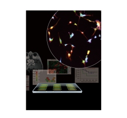 |
| Microfluidic imaging cytometry (MIC) platform |
Abstract:
Technology marks advance toward predictive and personalized medicine
By Jennifer Marcus
New diagnostic chip able to generate single-cell molecular 'fingerprints' for brain tumors
Los Angeles, CA | Posted on August 4th, 2010New technologies for the diagnosis of cancer are rapidly changing the clinical practice of oncology. As scientists learn more about the molecular basis of cancer, the development of new tools capable of multiple, inexpensive biomarker measurements on small samples of clinical tissue will become essential to the success of genetically informed and personalized cancer therapies.
Researchers at UCLA have now developed a microfluidic image cytometry (MIC) platform that can measure cell-signaling pathways in brain tumor samples at the single-cell level. The new technology combines the advantages of microfluidics and microscopy-based cell imaging.
The ability to make these in vitro molecular measurements, or "fingerprints," marks a new advance in molecular diagnostics that could ultimately help physicians predict patient prognosis and guide personalized treatment.
"The MIC is essentially a cancer diagnostic chip that can generate single-cell 'molecular fingerprints' for a small quantity of pathology samples, including brain tumor tissues," said Dr. Hsian-Rong Tseng, a UCLA associate professor of molecular and medical pharmacology and one of the leaders of the research. "We are exploring the use of the MIC for generating informative molecular fingerprints from rare populations of oncology samples � for example, tumor stem cells."
The research, which appears in the Aug. 1 issue of the journal Cancer Research, represents the teamwork of 35 co-authors from UCLA's Jonsson Comprehensive Cancer Center with expertise in surgery, pathology, cancer biology, bioinformatics and diagnostic devices.
Led by Tseng and Thomas Graeber, an assistant professor of molecular and medical pharmacology, both of whom are researchers at the Crump Institute for Molecular Imaging at the David Geffen School of Medicine at UCLA and the California NanoSystems Institute (CNSI) at UCLA, the team analyzed a panel of 19 human brain tumor biopsies to show the clinical application of the MIC platform to solid tumors.
The researchers also developed new bioinformatics � computational and statistical techniques and algorithms � that allowed them to process and analyze the data gleaned from the MIC platform's single-cell measurements.
"Because the measurements are at the single-cell level, computational algorithms are then used to organize and find patterns in the thousands of measurements," Graeber said. "These patterns relate to the growth signaling pathways active in the tumor that should be targeted in genetically informed or personalized anticancer therapies."
"The single-cell nature of the MIC brain tumor data presented an exciting and challenging opportunity," said Dr. Nicholas Graham, a postdoctoral scholar at the CNSI who worked out the data analysis. "To make sense of the data, we had to develop some new bioinformatics approaches that would preserve the power of single-cell analysis but allow for comparison between patients."
Molecular and medical pharmacology graduate researcher Michael Masterman-Smith approached the project as a translational cancer biologist.
"When we incorporated patient outcome data into our analyses and found that these 'biosignatures' clustered to reveal distinctive signaling phenomena that correlated with outcome, it got truly exciting," he said.
Microscale technology platforms are finding wide application in biological assays in which careful manipulation and measurement of limited sample amounts are required, and the new MIC platform is capable of making molecular measurements on small tumor samples provided by tumor resection and biopsy using as few as 1,000 to 3,000 cells, according to the researchers.
"The promise and attractiveness of this approach is the small amount of tissue needed for analysis in the face of increasing numbers of prognostic and predictive markers, and the possibility of quantifying tumor genetic heterogeneity," said Dr. William Yong, a Jonsson Cancer Center physician-scientist who led the pathology aspects of the research. "However, much work remains to validate this study with larger sample sizes and with more markers."
"We are excited about the possibility of using this method to investigate responses of individual tumors to potential therapeutics, as well as to enhance our knowledge about how they become resistant to therapies," said Dr. Harley Kornblum, a physician-scientist who studies brain tumor biology and is a member of both UCLA's Intellectual and Developmental Disabilities Research Center and the Johnson Cancer Center cell biology program area.
"Scientific, medical and engineering disciplines each have their own approach to problem-solving" said Dr. Jing Sun, a postdoctoral scholar at CNSI and an organic chemist. "For the innovative process to yield something useful, it must be faster, better, cheaper and � of course, with microscale technologies � smaller."
The researchers will next apply the new platform to larger cohorts of cancer patient samples and integrate the diagnostic approach into clinical trials of molecular therapies.
CytoScale Diagnostics has signed a letter of agreement regarding the technology mentioned in this paper.
This study was funded by the National Cancer Institute/ National Institutes of Health (NCI/NIH) and the National Institute of Neurological Disorders and Stroke (NINDS).
####
About UCLA's Jonsson Comprehensive Cancer Center
UCLA's Jonsson Comprehensive Cancer Center has more than 240 researchers and clinicians engaged in disease research, prevention, detection, control, treatment and education. One of the nation's largest comprehensive cancer centers, the Jonsson Center is dedicated to promoting research and translating basic science into leading-edge clinical studies. In July 2010, the Jonsson Cancer Center was named among the top 10 cancer centers nationwide by U.S. News & World Report, a ranking it has held for 10 of the last 11 years.
The California NanoSystems Institute at UCLA is an integrated research center operating jointly at UCLA and UC Santa Barbara whose mission is to foster interdisciplinary collaborations for discoveries in nanosystems and nanotechnology; train the next generation of scientists, educators and technology leaders; and facilitate partnerships with industry, fueling economic development and the social well-being of California, the United States and the world. The CNSI was established in 2000 with $100 million from the state of California and an additional $250 million in federal research grants and industry funding. At the institute, scientists in the areas of biology, chemistry, biochemistry, physics, mathematics, computational science and engineering are measuring, modifying and manipulating the building blocks of our world � atoms and molecules. These scientists benefit from an integrated laboratory culture enabling them to conduct dynamic research at the nanoscale, leading to significant breakthroughs in the areas of health, energy, the environment and information technology.
The UCLA Crump Institute for Molecular Imaging is a multidisciplinary collaborative of faculty, postdoctoral scholars and graduate students engaged in cutting-edge research in the fields of molecular diagnostics, microfluidics, systems biology and nanotechnology, with the aim of developing new technologies to observe, measure and understand biology in cells, tissues and living organisms through molecular imaging. The institute's ultimate goal is to provide the technology and science that will lead to a better understanding of the transition from health to disease at the molecular level and the development of new therapies to treat disease as part of a new era in molecular medicine.
For more information, please click here
Contacts:
Media Contacts
Jennifer Marcus,
310-267-4839
Copyright © UCLA's Jonsson Comprehensive Cancer Center
If you have a comment, please Contact us.Issuers of news releases, not 7th Wave, Inc. or Nanotechnology Now, are solely responsible for the accuracy of the content.
Bookmark:


| Related News Press |
News and information
![]() Quantum computer improves AI predictions April 17th, 2026
Quantum computer improves AI predictions April 17th, 2026
![]() Flexible sensor gains sensitivity under pressure April 17th, 2026
Flexible sensor gains sensitivity under pressure April 17th, 2026
![]() A reusable chip for particulate matter sensing April 17th, 2026
A reusable chip for particulate matter sensing April 17th, 2026
![]() Detecting vibrational quantum beating in the predissociation dynamics of SF6 using time-resolved photoelectron spectroscopy April 17th, 2026
Detecting vibrational quantum beating in the predissociation dynamics of SF6 using time-resolved photoelectron spectroscopy April 17th, 2026
Govt.-Legislation/Regulation/Funding/Policy
![]() Quantum computer improves AI predictions April 17th, 2026
Quantum computer improves AI predictions April 17th, 2026
![]() Metasurfaces smooth light to boost magnetic sensing precision January 30th, 2026
Metasurfaces smooth light to boost magnetic sensing precision January 30th, 2026
![]() New imaging approach transforms study of bacterial biofilms August 8th, 2025
New imaging approach transforms study of bacterial biofilms August 8th, 2025
Possible Futures
![]() A fundamentally new therapeutic approach to cystic fibrosis: Nanobody repairs cellular defect April 17th, 2026
A fundamentally new therapeutic approach to cystic fibrosis: Nanobody repairs cellular defect April 17th, 2026
![]() UC Irvine physicists discover method to reverse �quantum scrambling� : The work addresses the problem of information loss in quantum computing system April 17th, 2026
UC Irvine physicists discover method to reverse �quantum scrambling� : The work addresses the problem of information loss in quantum computing system April 17th, 2026
Academic/Education
![]() Rice University launches Rice Synthetic Biology Institute to improve lives January 12th, 2024
Rice University launches Rice Synthetic Biology Institute to improve lives January 12th, 2024
![]() Multi-institution, $4.6 million NSF grant to fund nanotechnology training September 9th, 2022
Multi-institution, $4.6 million NSF grant to fund nanotechnology training September 9th, 2022
Nanomedicine
![]() A fundamentally new therapeutic approach to cystic fibrosis: Nanobody repairs cellular defect April 17th, 2026
A fundamentally new therapeutic approach to cystic fibrosis: Nanobody repairs cellular defect April 17th, 2026
![]() New molecular technology targets tumors and simultaneously silences two �undruggable� cancer genes August 8th, 2025
New molecular technology targets tumors and simultaneously silences two �undruggable� cancer genes August 8th, 2025
![]() New imaging approach transforms study of bacterial biofilms August 8th, 2025
New imaging approach transforms study of bacterial biofilms August 8th, 2025
![]() Electrifying results shed light on graphene foam as a potential material for lab grown cartilage June 6th, 2025
Electrifying results shed light on graphene foam as a potential material for lab grown cartilage June 6th, 2025
Announcements
![]() A fundamentally new therapeutic approach to cystic fibrosis: Nanobody repairs cellular defect April 17th, 2026
A fundamentally new therapeutic approach to cystic fibrosis: Nanobody repairs cellular defect April 17th, 2026
![]() UC Irvine physicists discover method to reverse �quantum scrambling� : The work addresses the problem of information loss in quantum computing system April 17th, 2026
UC Irvine physicists discover method to reverse �quantum scrambling� : The work addresses the problem of information loss in quantum computing system April 17th, 2026
Nanobiotechnology
![]() A fundamentally new therapeutic approach to cystic fibrosis: Nanobody repairs cellular defect April 17th, 2026
A fundamentally new therapeutic approach to cystic fibrosis: Nanobody repairs cellular defect April 17th, 2026
![]() New molecular technology targets tumors and simultaneously silences two �undruggable� cancer genes August 8th, 2025
New molecular technology targets tumors and simultaneously silences two �undruggable� cancer genes August 8th, 2025
![]() New imaging approach transforms study of bacterial biofilms August 8th, 2025
New imaging approach transforms study of bacterial biofilms August 8th, 2025
![]() Electrifying results shed light on graphene foam as a potential material for lab grown cartilage June 6th, 2025
Electrifying results shed light on graphene foam as a potential material for lab grown cartilage June 6th, 2025
|
|
||
|
|
||
| The latest news from around the world, FREE | ||
|
|
||
|
|
||
| Premium Products | ||
|
|
||
|
Only the news you want to read!
Learn More |
||
|
|
||
|
Full-service, expert consulting
Learn More |
||
|
|
||