Home > Press > Wireless microgrippers grab living cells in 'biopsy' tests
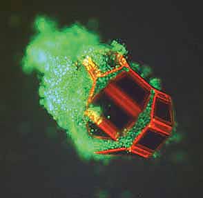 |
| This fluorescent micrograph features a single microgripper with live cells within its grasp. The tetherless microgripper was triggered to close and capture these live fibroblast cells with thermobiochemical cues. |
Abstract:
In experiments that pave the way for tiny mobile surgical tools activated by heat or chemicals, Johns Hopkins researchers have invented dust-particle-size devices that can be used to grab and remove living cells from hard-to-reach places without the need for electrical wires, tubes or batteries. Instead, the devices are actuated by thermal or biochemical signals.
Wireless microgrippers grab living cells in 'biopsy' tests
Baltimore, MD | Posted on January 12th, 2009The mass-producible microgrippers each measure approximately one-tenth of a millimeter in diameter. In lab tests, they have been used to perform a biopsy-like procedure on animal tissue placed at the end of a narrow tube. Experiments using the devices were reported in the online Early Edition of Proceedings of the National Academy of Sciences for the week of Jan. 12-16.
Although the devices will require further refinement before they can be used in humans, David H. Gracias, who supervised the project, said these thermobiochemically responsive, functional micro-tools represent a paradigm shift in engineering. "We've demonstrated tiny inexpensive tools that can be triggered en masse by nontoxic biochemicals," said Gracias, an assistant professor of chemical and biomolecular engineering in Johns Hopkins' Whiting School of Engineering. "This is an important first step toward creating a new set of biochemically responsive and perhaps even autonomous micro- and nanoscale surgical tools that could help doctors diagnose illnesses and administer treatment in a more efficient, less invasive way."
Today, doctors who wish to collect cells or manipulate a bit of tissue inside a patient's body often use tethered microgrippers connected to thin wires or tubes. But these tethers can make it difficult navigate the tool through tortuous or hard-to-reach locations. To eliminate this problem, the untethered grippers devised by Gracias' team contain gold-plated nickel, allowing them to be steered by magnets outside the body. "With this method, we were able to remotely move the microgrippers a relatively long distance over tissue without getting stuck, he said. "Additionally, the microgrippers are triggered to close and extricate cells from tissue when exposed to certain biochemicals or biologically relevant temperatures."
The microgripper design -- six three-jointed digits extended from a central "palm" -- resembles a crab. (In fact, the joint design was inspired by that of arthropod animals.) To fabricate the microgrippers in their initial flat position with all digits fully extended, the researchers employ photolithography, the same process used to make computer chips. When the tiny devices are inserted in the body and moved magnetically, the gold-plated nickel in the palm and digits will allow doctors to see and guide the grippers with medical imaging units such as an MRI or CT.
The microgrippers' grasping ability is rooted in the chemical composition of the joints embedded in the finger-like digits. These joints contain thin layers of chromium and copper with stress characteristics that would normally cause the digits to curl themselves closed like fingers clasping a baseball. But the researchers added a polymer resin, giving the joints rigidity to keep the fingers from closing.
When the microgrippers arrive at their destination, however, the researchers raise the temperature to 40 degrees C (or 104 degrees F, equivalent to a moderate fever in humans). This heat softens the polymer in the joints, causing the fingers to flex shut. The researchers also found an alternative method: Some nontoxic biological solutions can also weaken the polymer and cause the grippers to clamp down on their target.
In their lab experiments, the Johns Hopkins researchers used a microgripper, guided by a magnet, to grab and transport a dyed bead from among a group of colorless beads in a water solution. Team members also captured dozens of live animal cells from a cell mass at the end of a capillary tube. The cells were still alive 72 hours later, indicating the capture process did not injure them. Also, the microgrippers captured samples from relatively tough bovine bladder tissue.
The experiments showed that the tetherless microgripper concept is viable and has great potential for medical applications, the researchers said. Gracias' team is now working to overcome some remaining hurdles. As currently designed, each biologically compatible gripper can close on a target only once and cannot be reactivated to reopen and release its contents. (A similar device from the Gracias team, aimed at industrial micro-assembly applications, can be directed to both capture and release its load, but this requires chemicals that are not safe for patients. This pick-and-place microgripper was described in a recent article in the Journal of the American Chemical Society.)
Gracias, who also is affiliated with the Institute for NanoBioTechnology at Johns Hopkins, hopes to collaborate with medical researchers who can help to move the microgrippers closer to use as practical biopsy and drug delivery tools in humans. In September, he received a $1.5 million New Innovators Award from the National Institutes of Health. He plans to use the five-year grant to develop an entire mobile, biochemically responsive micro- and nanoscale surgical tool kit.
The lead author of the PNAS microgripper article was Timothy G. Leong, who was a doctoral student supervised by Gracias. In addition to Leong and Gracias, the paper's co-authors, all students supervised by Gracias at Johns Hopkins, were Christina L. Randall, a doctoral student in the Department of Biomedical Engineering; Brian R. Benson, a junior undergraduate supported by a Provost's Undergraduate Research Award; Noy Bassik, who is enrolled in an M.D./Ph.D. program involving the School of Medicine and the Department of Chemical and Biomolecular Engineering; and George M. Stern, a master's degree student in chemical and biomolecular engineering.
The Johns Hopkins Technology Transfer staff has obtained a provisional United States patent covering the team's inventions and is seeking international patent protection.
Funding for the research was provided by the National Science Foundation, the National Institutes of Health, and the Dreyfus and Beckman foundations.
Photos, illustrations and videos may be seen online at www.jhu.edu/news/home09/jan09/gracias.html and are available by contacting Phil Sneiderman.
####
For more information, please click here
Contacts:
Phil Sneiderman
443-287-9960
Copyright © Johns Hopkins University
If you have a comment, please Contact us.Issuers of news releases, not 7th Wave, Inc. or Nanotechnology Now, are solely responsible for the accuracy of the content.
Bookmark:


| Related Links |
![]() Department of Chemical and Biomolecular Engineering
Department of Chemical and Biomolecular Engineering
![]() "Tetherless Thermo-biochemically Actuated Microgrippers"
"Tetherless Thermo-biochemically Actuated Microgrippers"
![]() Journal of the American Chemical Society: "Pick-and-Place Using Chemically
Journal of the American Chemical Society: "Pick-and-Place Using Chemically
| Related News Press |
News and information
![]() Quantum computer improves AI predictions April 17th, 2026
Quantum computer improves AI predictions April 17th, 2026
![]() Flexible sensor gains sensitivity under pressure April 17th, 2026
Flexible sensor gains sensitivity under pressure April 17th, 2026
![]() A reusable chip for particulate matter sensing April 17th, 2026
A reusable chip for particulate matter sensing April 17th, 2026
![]() Detecting vibrational quantum beating in the predissociation dynamics of SF6 using time-resolved photoelectron spectroscopy April 17th, 2026
Detecting vibrational quantum beating in the predissociation dynamics of SF6 using time-resolved photoelectron spectroscopy April 17th, 2026
Nanomedicine
![]() A fundamentally new therapeutic approach to cystic fibrosis: Nanobody repairs cellular defect April 17th, 2026
A fundamentally new therapeutic approach to cystic fibrosis: Nanobody repairs cellular defect April 17th, 2026
![]() New molecular technology targets tumors and simultaneously silences two �undruggable� cancer genes August 8th, 2025
New molecular technology targets tumors and simultaneously silences two �undruggable� cancer genes August 8th, 2025
![]() New imaging approach transforms study of bacterial biofilms August 8th, 2025
New imaging approach transforms study of bacterial biofilms August 8th, 2025
![]() Electrifying results shed light on graphene foam as a potential material for lab grown cartilage June 6th, 2025
Electrifying results shed light on graphene foam as a potential material for lab grown cartilage June 6th, 2025
Discoveries
![]() Quantum computer improves AI predictions April 17th, 2026
Quantum computer improves AI predictions April 17th, 2026
![]() Flexible sensor gains sensitivity under pressure April 17th, 2026
Flexible sensor gains sensitivity under pressure April 17th, 2026
![]() A reusable chip for particulate matter sensing April 17th, 2026
A reusable chip for particulate matter sensing April 17th, 2026
![]() Detecting vibrational quantum beating in the predissociation dynamics of SF6 using time-resolved photoelectron spectroscopy April 17th, 2026
Detecting vibrational quantum beating in the predissociation dynamics of SF6 using time-resolved photoelectron spectroscopy April 17th, 2026
Announcements
![]() A fundamentally new therapeutic approach to cystic fibrosis: Nanobody repairs cellular defect April 17th, 2026
A fundamentally new therapeutic approach to cystic fibrosis: Nanobody repairs cellular defect April 17th, 2026
![]() UC Irvine physicists discover method to reverse �quantum scrambling� : The work addresses the problem of information loss in quantum computing system April 17th, 2026
UC Irvine physicists discover method to reverse �quantum scrambling� : The work addresses the problem of information loss in quantum computing system April 17th, 2026
|
|
||
|
|
||
| The latest news from around the world, FREE | ||
|
|
||
|
|
||
| Premium Products | ||
|
|
||
|
Only the news you want to read!
Learn More |
||
|
|
||
|
Full-service, expert consulting
Learn More |
||
|
|
||