Home > Nanotechnology Columns > Cristian Orfescu > NanoArt 2007 International Online Competition - Part I
 |
Cris Orfescu Founder NanoArt21 |
Abstract:
The voting process for the NanoArt 2007 International Online Competition is over. 121 works were submited by 37 nanoartists from 13 countries and the public voting was open during a period of 3 months. Congratulations to all participating artists! The first 10 artists will be featured in a two-parts article.
April 16th, 2008
NanoArt 2007 International Online Competition - Part I
For a couple of years, I organize an international online competition for NanoArt, a new artistic discipline, a logic follow-up of the Nanotechnology development.
The artists and scientists are encouraged to participate with their own images as long as these visualize micro or nano structures. However, because NanoArt is still in incipient stage, NanoArt21 ( http://nanoart21.org ) provided for competitors 3 high resolution monochromatic electron scans, to choose from. The artists who desired to participate but did not have at the contest time the possibility to create NanoArt from scratch and to get their own images, were offered the option to alter the seed images in any artistic way to finish the artistic-scientific process and create NanoArt works.
This year, 121 artworks were submited by 37 nanoartists from 13 countries and the public voting was open during a period of 3 months.
As I mentioned in a previous article, several techniques have been used to morph artistically the micro or nanostructures images generated in the advanced microscopes.
On the first place came Teresa Majerus from Luxembourg ( http://www.art-of-kreska.eu )
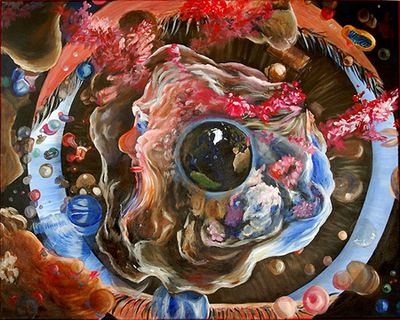 |
| Eye of science, by Teresa Majerus, acrylics on canvas |
"Rather than use the nanoflower seed image as a basis for the further electronic manipulation, I decided to use it as inspiration for an acrylic painting on canvas. The painted canvas shows a view of the nanoworld seen from the particle perspective. We can clearly recognize the eye of the scientist looking via microscope onto the complex but beautiful structure. In her/his iris our world is reflected, which in the same time appears to be the core of nanospace. The model of the atom plays here the role of an engine. The story of progress started ages ago. To remind us about great minds and troubles of education in the past, man and woman (left and right), were placed on the opposite sides of the unified mass. They are very distinctive, individual. Maybe their approach of solving the problems is sometimes different, but if they consolidate their forces, as it is symbolized here by their unification within one solid body, they are the busters of development. The sundial in the dress of the woman reminds us of the passing time. Every story has its beginning, but where starts nanoscience? The simplest explanation of how microscopes work is referring to a telescope. Therefore, as a tribute to the excellent scientist, the Newton's telescope can be found on the picture. The things sometimes seem to us more complex then they are. Who was not in the situation when the answer to our riddle seemed to be an enigma, whereas it was lying just in front of us? That is exactly what the monkey represents in my creation. This recalls us Darwin, and his theory of evolution. The biological element (mitochondrion) somehow rounds up this story. There are few more things worth noting. Let us go from the beard of the man to the left lower corner. One of the hairs continues like a thread (maybe it is a DNA carrying important information), goes via a needle (kind of selection process) and ends onto twisted together coil. This is a beginning of creation, which is a continuum of embryonic and fetal life. In this course, the human curiosity appears, in order to know about health and gender. These questions are answered by ultrasound examination and we see hope, the next generation of a great scientist. Finally, the work of the scientists is awarded and champagne is served.", says Majerus, describing her artwork.
On the second place came Chris Robinson, Associate Chair and Professor, Studio Art: Visual Computing, 3D Studies at the University of South Carolina, USA ( http://www.cas.sc.edu/art/Faculty/robinsonc/robinsonc.htm )
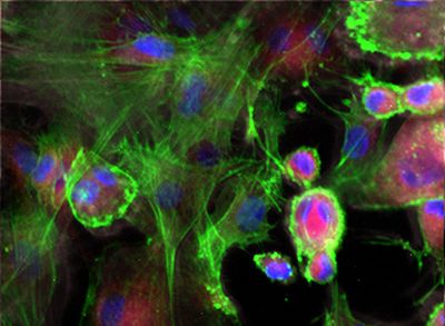 |
| Untitled (Transfect), by Chris Robinson |
Here is the description of Robinson's work: "This confocal image is of transfected neonatal rat cardiac fibroblasts expressing fluorescently-tagged DDR2 (Discoidin Domain Receptor 2). The cells are in a collagen thick film, which simulates the extracellular environment. DDR2 is a transmembrane protein, which is thought to be involved in sensing the cell's local mechanical environment and signaling to the cell the need for remodeling. The blue stain shows the cell nuclei, the green stain shows the actin filaments of the cell cytoskeleton, and the red punctate staining shows the spatial location of the DDR2. (S. Baxter - Mech. Eng., E. Goldsmith - Cell Biology, C. Murphy -Chemistry, C. Robinson - Art; University of South Carolina NanoCenter)."
An excellent digital artist, specialized in fractal art, Renata Spiazzi came in the 3rd place. Here is the link to the artist's website: http://www.spiazzi.com
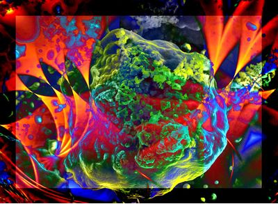 |
| Nano Depths, by Renata Spiazzi, fractals |
Spiazzi's work was created with several overlappings of fractals and the seed image of the nanostructure. The programs used were Apophysis, Ultrafractal and Photoshop plus filters from the KPT group.
Bookmark:


|
|
||
|
|
||
| The latest news from around the world, FREE | ||
|
|
||
|
|
||
| Premium Products | ||
|
|
||
|
Only the news you want to read!
Learn More |
||
|
|
||
|
Full-service, expert consulting
Learn More |
||
|
|
||