Home > Press > Imaging single spine structural plasticity at the nanoscale level: Researchers at the Max Planck Florida Institute for Neuroscience (MPFI) have developed a new imaging technique capable of visualizing the dynamically changing structure of dendritic spines with unprecedented resol
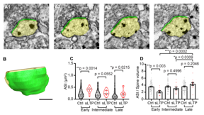 |
| A, B, Serial images of an example spine (A) and 3D reconstruction of the example spine (B) with spine head shown in yellow and ASI shown in green. Scale bar, 200 nm. C, ASI area size at early, intermediate, and late phases of sLTP for sLTP and control (Ctrl) spines. Welch�s t test was used on log-transformed data. D, Ratio of ASI area to spine volume at early, intermediate, and late phases of sLTP. CREDIT Max Planck Florida Institute for Neuroscience |
Abstract:
For most, the relentless snapping of camera shutters is an all too familiar sound associated with trips and vacations. When venturing to a new place, travelers everywhere are constantly on the search for that picture-perfect, Instagram worthy shot. Persevering through many takes, amateur photographers fight blurred backgrounds, closed eyes, and photo-bombing passersby all in search of that ever-elusive perfect picture.
Imaging single spine structural plasticity at the nanoscale level: Researchers at the Max Planck Florida Institute for Neuroscience (MPFI) have developed a new imaging technique capable of visualizing the dynamically changing structure of dendritic spines with unprecedented resol
West Palm Beach, FL | Posted on September 3rd, 2021As it turns out, neuroscientists are very similar to travelers in this regard, constantly developing and practicing new ways to take perfect, crystal-clear images. But instead of picturesque natural backdrops or striking city scenes, neuroscientists are interested in detailed snapshots of brain cells and their small-scale structures.
The Yasuda Lab at MPFI is incredibly well versed in small-scale structures of the brain, focused on studying the dynamic changes to tiny synaptic compartments called dendritic spines. Robust changes in spine structure known as structural plasticity, allow synapses to robustly modulate their connection strength. By doing so, cells in the brain can actively strengthen important connections and weaken those that are less needed. This process is thought to underlie how we learn and remember. But revealing the fine structures of spines in detail during such a dynamic process is a difficult undertaking. Until recently, imaging methodologies lacked the capabilities to do so.
In a recent publication in The Journal of Neuroscience, researchers in the Yasuda Lab have developed a powerful new imaging strategy capable of visualizing the fine, ultrastructural changes to dendritic spines during structural plasticity. By modifying and building off an established imaging technique known as correlative light and electron microscopy (CLEM), MPFI scientists have harnessed the best that both imaging modalities can provide.
�Dendritic spines are such small-scale neuronal compartments, that it�s difficult to get an accurate picture of what�s actually occurring in terms of structural changes using traditional imaging methods,� explains Dr. Ryohei Yasuda, Scientific Director at MPFI. �Using more standard optical techniques like 2-photon microscopy, dendritic spines look like smooth spheres. In actuality, we know from using more powerful imaging methods, like electron microscopy, that the actual size and shape of spines are far more complex. So, we were interested in learning what changes occur during the various stages of structural plasticity, at a resolution where we could take a deeper look at the spine�s complexity.�
The MPFI team first induced structural plasticity in single dendritic spines using 2-photon optical microscopy and glutamate uncaging. The induced spine was then fixed in time at one of three distinct timepoints, representing the major stages of structural plasticity. In close collaboration with MPFI�s Electron Microscopy (EM) Core, brain tissue samples containing the stimulated spines were cut into ultra-thin sections using a specialized device called ATUMtome. These sections were then re-imaged using the extreme resolving power of the Electron Microscope to reveal the ultrastructural details and reconstruct accurate pictures of the spine�s complex topography.
�When we started this project, our goal was to see if it was even possible to collect spines at various stages of structural plasticity, successfully relocate them, and resolve their ultrastructure using EM,� describes Ye Sun, Ph.D., former Graduate Student in the Yasuda Lab and first author of the publication. �Single, spine-specific forms of structural plasticity have never been imaged in this way before. Dr. Naomi kamasawa, Head of MPFI�s EM Core, was instrumental in helping to establish and optimize our EM workflow for the project.�
Examining the reconstructed spine images, the MPFI team noticed unique changes to a protein-rich region of dendritic spines, called the postsynaptic density (PSD). This region is critically important for the spine, implicated in regulating synaptic strength and plasticity. MPFI researchers found that compared to control spines, the area and size of the PSD region was significantly greater in spines that underwent structural plasticity. PSD growth in these spines occurred on a slower timescale, needing hours to reach its maximal change. Interestingly while growth was on a slower scale, PSD structure in stimulated spines reorganized at a rapid pace. After the induction of structural plasticity, PSD complexity immediately increased, dramatically transforming in shape and structural features.
�Our imaging strategy synergizes the best of both optical and EM microscopies, allowing us to study spine structural changes never before seen in nanoscale resolution,� notes Dr. Yasuda. �For the future, our lab is interested in using this new protocol in combination with advanced molecular techniques, such as SLENDR, to study individual protein dynamics in tandem with finely detailed structural changes during spine structural plasticity.
####
For more information, please click here
Contacts:
Helena Decker
Max Planck Florida Institute for Neuroscience
Office: 561-972-9253
Copyright © Max Planck Florida Institute for Neuroscience
If you have a comment, please Contact us.Issuers of news releases, not 7th Wave, Inc. or Nanotechnology Now, are solely responsible for the accuracy of the content.
Bookmark:


| Related Links |
| Related News Press |
News and information
![]() Simulating magnetization in a Heisenberg quantum spin chain April 5th, 2024
Simulating magnetization in a Heisenberg quantum spin chain April 5th, 2024
![]() NRL charters Navy�s quantum inertial navigation path to reduce drift April 5th, 2024
NRL charters Navy�s quantum inertial navigation path to reduce drift April 5th, 2024
![]() Discovery points path to flash-like memory for storing qubits: Rice find could hasten development of nonvolatile quantum memory April 5th, 2024
Discovery points path to flash-like memory for storing qubits: Rice find could hasten development of nonvolatile quantum memory April 5th, 2024
Imaging
![]() Nanoscale CL thermometry with lanthanide-doped heavy-metal oxide in TEM March 8th, 2024
Nanoscale CL thermometry with lanthanide-doped heavy-metal oxide in TEM March 8th, 2024
![]() The USTC realizes In situ electron paramagnetic resonance spectroscopy using single nanodiamond sensors November 3rd, 2023
The USTC realizes In situ electron paramagnetic resonance spectroscopy using single nanodiamond sensors November 3rd, 2023
![]() Observation of left and right at nanoscale with optical force October 6th, 2023
Observation of left and right at nanoscale with optical force October 6th, 2023
Possible Futures
![]() Discovery points path to flash-like memory for storing qubits: Rice find could hasten development of nonvolatile quantum memory April 5th, 2024
Discovery points path to flash-like memory for storing qubits: Rice find could hasten development of nonvolatile quantum memory April 5th, 2024
![]() With VECSELs towards the quantum internet Fraunhofer: IAF achieves record output power with VECSEL for quantum frequency converters April 5th, 2024
With VECSELs towards the quantum internet Fraunhofer: IAF achieves record output power with VECSEL for quantum frequency converters April 5th, 2024
Nanomedicine
![]() New micromaterial releases nanoparticles that selectively destroy cancer cells April 5th, 2024
New micromaterial releases nanoparticles that selectively destroy cancer cells April 5th, 2024
![]() Good as gold - improving infectious disease testing with gold nanoparticles April 5th, 2024
Good as gold - improving infectious disease testing with gold nanoparticles April 5th, 2024
![]() Researchers develop artificial building blocks of life March 8th, 2024
Researchers develop artificial building blocks of life March 8th, 2024
Discoveries
![]() Chemical reactions can scramble quantum information as well as black holes April 5th, 2024
Chemical reactions can scramble quantum information as well as black holes April 5th, 2024
![]() New micromaterial releases nanoparticles that selectively destroy cancer cells April 5th, 2024
New micromaterial releases nanoparticles that selectively destroy cancer cells April 5th, 2024
![]() Utilizing palladium for addressing contact issues of buried oxide thin film transistors April 5th, 2024
Utilizing palladium for addressing contact issues of buried oxide thin film transistors April 5th, 2024
Announcements
![]() NRL charters Navy�s quantum inertial navigation path to reduce drift April 5th, 2024
NRL charters Navy�s quantum inertial navigation path to reduce drift April 5th, 2024
![]() Discovery points path to flash-like memory for storing qubits: Rice find could hasten development of nonvolatile quantum memory April 5th, 2024
Discovery points path to flash-like memory for storing qubits: Rice find could hasten development of nonvolatile quantum memory April 5th, 2024
Interviews/Book Reviews/Essays/Reports/Podcasts/Journals/White papers/Posters
![]() Simulating magnetization in a Heisenberg quantum spin chain April 5th, 2024
Simulating magnetization in a Heisenberg quantum spin chain April 5th, 2024
![]() Discovery points path to flash-like memory for storing qubits: Rice find could hasten development of nonvolatile quantum memory April 5th, 2024
Discovery points path to flash-like memory for storing qubits: Rice find could hasten development of nonvolatile quantum memory April 5th, 2024
Tools
![]() Ferroelectrically modulate the Fermi level of graphene oxide to enhance SERS response November 3rd, 2023
Ferroelectrically modulate the Fermi level of graphene oxide to enhance SERS response November 3rd, 2023
![]() The USTC realizes In situ electron paramagnetic resonance spectroscopy using single nanodiamond sensors November 3rd, 2023
The USTC realizes In situ electron paramagnetic resonance spectroscopy using single nanodiamond sensors November 3rd, 2023
Nanobiotechnology
![]() New micromaterial releases nanoparticles that selectively destroy cancer cells April 5th, 2024
New micromaterial releases nanoparticles that selectively destroy cancer cells April 5th, 2024
![]() Good as gold - improving infectious disease testing with gold nanoparticles April 5th, 2024
Good as gold - improving infectious disease testing with gold nanoparticles April 5th, 2024
![]() Researchers develop artificial building blocks of life March 8th, 2024
Researchers develop artificial building blocks of life March 8th, 2024
|
|
||
|
|
||
| The latest news from around the world, FREE | ||
|
|
||
|
|
||
| Premium Products | ||
|
|
||
|
Only the news you want to read!
Learn More |
||
|
|
||
|
Full-service, expert consulting
Learn More |
||
|
|
||