Home > Press > Membrane nano-tomography in living cells: Label-free evanescent microscopy enables full-field and real-time tracking of membrane processes without signal fading and cell perturbation
 |
Abstract:
Membranes play a pivotal role in numerous cell mechanisms, in particular for internalization, adhesion and motility studies. In terms of optical imaging of the membrane, special configurations are needed to remove the light coming from the inner part of the cell. French scientists now show that through-the-objective evanescent microscopy (epi-EM) is a powerful technique to image membranes in living cells.
Membrane nano-tomography in living cells: Label-free evanescent microscopy enables full-field and real-time tracking of membrane processes without signal fading and cell perturbation
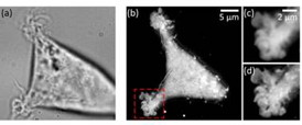
Paris, France | Posted on December 5th, 2014In label-free evanescent microscopy (EM), configurations similar to total internal reflection fluorescence (TIRF) have been proposed: prism-based or through-the-objective. However, in the latter case, these evanescent techniques have not spread much, with a relative preference for the prism-based configuration also called total internal reflection microscopy (TIRM). The team led by Pierre Bon chose the through-the-objective based configuration (epi-EM), which enabled super-axially resolved tomographic reconstruction of the basal membrane of label-free living cells. The implementation of epi-EM only required an easy to settle illumination/collection scheme on a standard inverted microscope. Only a high-NA objective (NAobj > 1.33) was needed for living biological sample studies and a spatial filter on the epi-illumination arm in order to reject under-critical angle illumination.
Either bead calibration or a multilayer Fresnel model could be used to retrieve nanometric position. Based on a multilayer Fresnel model, the team was able to retrieve the membrane/interface distance with 10 nm precision. The researchers applied this nano-axial tomography to retrieve quantitative information on invagination dynamics of living cell membranes. They studied the membrane elevation map of living cells (Wt HEK-293) during 15 minutes at one frame per second without perturbing the sample.
The results demonstrate that epi-EM gives easily access to axially super-resolved images of unlabeled microscopic samples with almost no microscope modification, and at least a doubled lateral resolution compared to classical TIRM. A study can be of any duration as the signal level is not sensitive to any fluorophore stability dependence and the photoxicity is very low as barely any light is absorbed by the sample. The scientists are convinced that this technique will be useful for cell motility and adhesion studies when the sample cannot be modified (ex. stem cells) or when very fast and/or long studies are required.
(Text contributed by K. Maedefessel-Herrmann)
####
About Journal of Biophotonics
Journal of Biophotonics publishes cutting edge research on interactions between light and biological material. The journal is highly interdisciplinary, covering research in the fields of physics, chemistry, biology and medicine. The scope extends from basic research to clinical applications. Connecting scientists who try to understand basic biological processes using light as a diagnostic and therapeutic tool, the journal offers a platform where the physicist communicates with the biologist and where the clinical practitioner learns about the latest tools for diagnosis of diseases. JBP offers fast publication times: down to 20 days from acceptance to publication. Impact Factor 2013: 3.856
For more information, please click here
Contacts:
Regina Hagen
Journal Publishing Manager
JBP
Copyright © Journal of Biophotonics
If you have a comment, please Contact us.Issuers of news releases, not 7th Wave, Inc. or Nanotechnology Now, are solely responsible for the accuracy of the content.
Bookmark:


| Related Links |
| Related News Press |
News and information
![]() Simulating magnetization in a Heisenberg quantum spin chain April 5th, 2024
Simulating magnetization in a Heisenberg quantum spin chain April 5th, 2024
![]() NRL charters Navy�s quantum inertial navigation path to reduce drift April 5th, 2024
NRL charters Navy�s quantum inertial navigation path to reduce drift April 5th, 2024
![]() Discovery points path to flash-like memory for storing qubits: Rice find could hasten development of nonvolatile quantum memory April 5th, 2024
Discovery points path to flash-like memory for storing qubits: Rice find could hasten development of nonvolatile quantum memory April 5th, 2024
Imaging
![]() Nanoscale CL thermometry with lanthanide-doped heavy-metal oxide in TEM March 8th, 2024
Nanoscale CL thermometry with lanthanide-doped heavy-metal oxide in TEM March 8th, 2024
![]() The USTC realizes In situ electron paramagnetic resonance spectroscopy using single nanodiamond sensors November 3rd, 2023
The USTC realizes In situ electron paramagnetic resonance spectroscopy using single nanodiamond sensors November 3rd, 2023
![]() Observation of left and right at nanoscale with optical force October 6th, 2023
Observation of left and right at nanoscale with optical force October 6th, 2023
Nanomedicine
![]() New micromaterial releases nanoparticles that selectively destroy cancer cells April 5th, 2024
New micromaterial releases nanoparticles that selectively destroy cancer cells April 5th, 2024
![]() Good as gold - improving infectious disease testing with gold nanoparticles April 5th, 2024
Good as gold - improving infectious disease testing with gold nanoparticles April 5th, 2024
![]() Researchers develop artificial building blocks of life March 8th, 2024
Researchers develop artificial building blocks of life March 8th, 2024
Discoveries
![]() Chemical reactions can scramble quantum information as well as black holes April 5th, 2024
Chemical reactions can scramble quantum information as well as black holes April 5th, 2024
![]() New micromaterial releases nanoparticles that selectively destroy cancer cells April 5th, 2024
New micromaterial releases nanoparticles that selectively destroy cancer cells April 5th, 2024
![]() Utilizing palladium for addressing contact issues of buried oxide thin film transistors April 5th, 2024
Utilizing palladium for addressing contact issues of buried oxide thin film transistors April 5th, 2024
Announcements
![]() NRL charters Navy�s quantum inertial navigation path to reduce drift April 5th, 2024
NRL charters Navy�s quantum inertial navigation path to reduce drift April 5th, 2024
![]() Discovery points path to flash-like memory for storing qubits: Rice find could hasten development of nonvolatile quantum memory April 5th, 2024
Discovery points path to flash-like memory for storing qubits: Rice find could hasten development of nonvolatile quantum memory April 5th, 2024
Interviews/Book Reviews/Essays/Reports/Podcasts/Journals/White papers/Posters
![]() Simulating magnetization in a Heisenberg quantum spin chain April 5th, 2024
Simulating magnetization in a Heisenberg quantum spin chain April 5th, 2024
![]() Discovery points path to flash-like memory for storing qubits: Rice find could hasten development of nonvolatile quantum memory April 5th, 2024
Discovery points path to flash-like memory for storing qubits: Rice find could hasten development of nonvolatile quantum memory April 5th, 2024
Tools
![]() Ferroelectrically modulate the Fermi level of graphene oxide to enhance SERS response November 3rd, 2023
Ferroelectrically modulate the Fermi level of graphene oxide to enhance SERS response November 3rd, 2023
![]() The USTC realizes In situ electron paramagnetic resonance spectroscopy using single nanodiamond sensors November 3rd, 2023
The USTC realizes In situ electron paramagnetic resonance spectroscopy using single nanodiamond sensors November 3rd, 2023
|
|
||
|
|
||
| The latest news from around the world, FREE | ||
|
|
||
|
|
||
| Premium Products | ||
|
|
||
|
Only the news you want to read!
Learn More |
||
|
|
||
|
Full-service, expert consulting
Learn More |
||
|
|
||