Home > Press > All Wrapped Up: Researcher's Graphene Cloak Protects Bacteria, Leading to Better Images
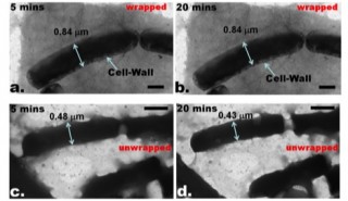 |
| This image shows the differences between bacteria that has been wrapped with graphene and bacteria that has not been wrapped. The images were taken after five minutes and after 20 minutes under an electron microscope, showing that the wrapped bacteria maintains a clear image. |
Abstract:
It's a cloak that surpasses all others: a microscopic carbon cloak made of graphene that could change the way bacteria and other cells are imaged.
All Wrapped Up: Researcher's Graphene Cloak Protects Bacteria, Leading to Better Images
Manhattan, KS | Posted on March 16th, 2011Vikas Berry, assistant professor of chemical engineering at Kansas State University, and his research team are wrapping bacteria with graphene to address current challenges with imaging bacteria under electron microscopes. Berry's method creates a carbon cloak that protects the bacteria, allowing them to be imaged at their natural size and increasing the image's resolution.
Graphene is a form of carbon that is only one atom thick, giving it several important properties: it's impermeable, it's the strongest nanomaterial, it's optically transparent and it has high thermal conductance.
"Graphene is the next-generation material," Berry said. "Although only an atom thick, graphene does not allow even the smallest of molecules to pass through. Furthermore, it's strong and highly flexible so it can conform to any shape."
Berry's team has been researching graphene for three years, and Berry recently saw a connection between graphene and cell imaging research. Because graphene is impermeable, he decided to use the material to preserve the size of bacterial cells imaged under high-vacuum electron microscopes.
The research results appear in the paper "Impermeable Graphenic Encasement of Bacteria," which was published in a recent issue of Nano Letters, a monthly scientific journal published by the American Chemical Society. The team's preliminary research appeared in Nature News in 2010.
The current challenge with cell imaging occurs when scientists use electron microscopes to image bacterial cells. Because these microscopes require a high vacuum, they remove water from the cells. Biological cells contain 70 to 80 percent water, and the result is a severely shrunk cell. As a result, it is challenging to obtain an accurate image of the cells and their components in their natural state.
But Berry and his team created a solution to the imaging challenge by applying graphene. The graphene acts as an impermeable cloak around the bacteria so that the cells retain water and don't shrink under the high vacuum of electron microscopes. This provides a microscopic image of the cell at its natural size.
The carbon cloaks can be wrapped around the bacteria using two methods. The first method involves putting a sheet of graphene on top of the bacteria, much like covering up with a bed sheet. The other method involves wrapping the bacteria with a graphene solution, where the graphene sheets swaddle the bacteria. In both cases the graphene sheets were functionalized with a protein to enhance binding with the bacterial cell wall.
Under the high vacuum of an electron microscope, the wrapped bacteria did not change in size for 30 minutes, giving scientists enough time to observe them. This is a direct result of the high strength and impermeability of the graphene cloak, Berry said.
Graphene's other extraordinary properties enhance the imaging resolution in microscopy. Its electron-transparency enables a clean imaging of the cells. Since graphene is a good conductor of heat and electricity, the local electronic-charging and heating is conducted off the graphene cloak, giving a clear view of the bacterial cell well. Unwrapped bacterial cells appear dark with an indistinguishable cell wall.
"Uniquely, graphene has all the properties needed to image bacteria at high resolutions," Berry said. "The project provides a very simple route to image samples in their native wet state."
The process has potential to influence future research. Scientists have always had trouble observing liquid samples under electron microscopes, but using carbon cloaks could allow them to image wet samples in a vacuum. Graphene's strong and impermeable characteristics ensure that wrapped cells can be easily imaged without degrading them. Berry said it might be possible in the future to use graphene to keep bacterium alive in a vacuum while observing its biochemistry under a microscope.
The research also paves the way for enhanced protein microscopy. Proteins act differently when they are dry and when they are in an aqueous solution. So far most protein studies have been conducted in dry phases, but Berry's research may allow proteins to be observed more in aqueous environments.
"This research could be the point of evolution for processing of sensitive samples with graphene to achieve enhanced imaging," Berry said.
Other researchers involved in the project include Daniel Boyle, research assistant professor in biology; Nihar Mohanty, doctoral student in chemical engineering, India; Ashvin Nagaraja, former master's student in electrical engineering; and Monica Fahrenholtz, a May 2010 chemical engineering graduate from Clearwater.
Berry also recently received a five-year, $400,000 CAREER award to study graphene quantum dots, which are ultrasmall sheets of carbon atoms.
####
For more information, please click here
Contacts:
Vikas Berry
785-532-5519
Copyright © Kansas State University
If you have a comment, please Contact us.Issuers of news releases, not 7th Wave, Inc. or Nanotechnology Now, are solely responsible for the accuracy of the content.
Bookmark:


| Related News Press |
News and information
![]() Simulating magnetization in a Heisenberg quantum spin chain April 5th, 2024
Simulating magnetization in a Heisenberg quantum spin chain April 5th, 2024
![]() NRL charters Navy�s quantum inertial navigation path to reduce drift April 5th, 2024
NRL charters Navy�s quantum inertial navigation path to reduce drift April 5th, 2024
![]() Discovery points path to flash-like memory for storing qubits: Rice find could hasten development of nonvolatile quantum memory April 5th, 2024
Discovery points path to flash-like memory for storing qubits: Rice find could hasten development of nonvolatile quantum memory April 5th, 2024
Imaging
![]() Nanoscale CL thermometry with lanthanide-doped heavy-metal oxide in TEM March 8th, 2024
Nanoscale CL thermometry with lanthanide-doped heavy-metal oxide in TEM March 8th, 2024
Graphene/ Graphite
![]() NRL discovers two-dimensional waveguides February 16th, 2024
NRL discovers two-dimensional waveguides February 16th, 2024
Discoveries
![]() Chemical reactions can scramble quantum information as well as black holes April 5th, 2024
Chemical reactions can scramble quantum information as well as black holes April 5th, 2024
![]() New micromaterial releases nanoparticles that selectively destroy cancer cells April 5th, 2024
New micromaterial releases nanoparticles that selectively destroy cancer cells April 5th, 2024
![]() Utilizing palladium for addressing contact issues of buried oxide thin film transistors April 5th, 2024
Utilizing palladium for addressing contact issues of buried oxide thin film transistors April 5th, 2024
Announcements
![]() NRL charters Navy�s quantum inertial navigation path to reduce drift April 5th, 2024
NRL charters Navy�s quantum inertial navigation path to reduce drift April 5th, 2024
![]() Discovery points path to flash-like memory for storing qubits: Rice find could hasten development of nonvolatile quantum memory April 5th, 2024
Discovery points path to flash-like memory for storing qubits: Rice find could hasten development of nonvolatile quantum memory April 5th, 2024
|
|
||
|
|
||
| The latest news from around the world, FREE | ||
|
|
||
|
|
||
| Premium Products | ||
|
|
||
|
Only the news you want to read!
Learn More |
||
|
|
||
|
Full-service, expert consulting
Learn More |
||
|
|
||