Home > Press > New technique takes a big step in examination of small structures
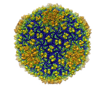 |
| Shown is an image of bacteriophage Epsilon15 studied by Wen Jiang, an assistant professor of biological sciences at Purdue. The bacteriophage is shown at a resolution of 4.5 angstrom - the highest resolution achieved for a living organism of this size. (Graphic/Wen Jiang lab) |
Abstract:
A team led by a Purdue University researcher has achieved images of a virus in detail two times greater than had previously been achieved.
New technique takes a big step in examination of small structures
WEST LAFAYETTE, IN | Posted on March 6th, 2008Wen Jiang, an assistant professor of biological sciences at Purdue, led a research team that used the emerging technique of single-particle electron cryomicroscopy to capture a three-dimensional image of a virus at a resolution of 4.5 angstroms. Approximately 1 million angstroms would equal the diameter of a human hair.
"This is one of the first projects to refine the technique to the point of near atomic-level resolution," said Jiang, who also is a member of Purdue's structural biology group. "This breaks a threshold and allows us to now see a whole new level of detail in the structure. This is the highest resolution ever achieved for a living organism of this size."
Details of the structure of a virus provide valuable information for development of disease treatments, he said.
"If we understand the system - how the virus particles assemble and how they infect a host cell - it will greatly improve our ability to design a treatment," Jiang said. "Structural biologists perform the basic science and provide information to help those working on the clinical aspects."
A paper detailing the work was published in the Feb. 28 issue of Nature.
Roger Hendrix, a professor of biological sciences at the University of Pittsburgh, said what is learned about viruses can be applied to many other biological systems.
"Understanding the proteins that create the structure of a virus gives us insight into the tiny biological machines found throughout our bodies," he said. "Getting to 4.5 angstrom using this technique is a watershed of sorts because it is the first time we can actually trace the polypeptide chain - the backbone of proteins. Now we can see the tiny gears and levers that allow the proteins to move and interact as they carry out their intricate biological roles."
The imaging technique, called cryo-EM, has the added benefit of maintaining the sample being studied in a state very similar to its natural environment. Other imaging techniques used regularly, such as X-ray crystallography, require the sample be manipulated.
"This method offers a new approach for modeling the structure of proteins in other macromolecular assemblies, such as DNA, at near-native states," Jiang said. "The sample is purified in a solution that is very similar to the environment that would be found in a host cell. It is as if the virus is frozen in glass and it is alive and infectious while we examine it."
In addition to Jiang, Matthew L. Baker, Joanita Jakana and Wah Chiu from Baylor College of Medicine, and Peter R. Weigele and Jonathan King from Massachusetts Institute of Technology worked on the project, which was funded by the National Institutes of Health and the National Science Foundation.
The team obtained a three-dimensional map of the capsid, or protein shell, of the epsilon15 bacteriophage, a virus that infects bacteria and is a member of a family of viruses that are the most abundant life forms on Earth, Jiang said.
Other methods of determining the structure could not be used for this family of virus. None had been successfully crystallized, and the complexity of members of this family had prevented evaluation through the genome sequence alone.
"This demonstration shows that cryo-EM is doable and is a major step in reaching the full potential of this technique," he said. "The goal is to have it reach a 3 to 4 angstrom resolution, which would allow us to clearly see the amino acids that make up a protein."
In electron microscopy, a beam of electrons takes the place of the light beam used in a conventional microscope. The use of electrons instead of light allows the microscope to "see" in much greater detail.
Cryo-EM cools specimens to temperatures well below the freezing point of water. This decreases damage from the electron beam and allows the specimens to be examined for a longer period of time. Longer exposure time allows for sharper, more detailed images.
Researchers using cryo-EM had obtained images at a resolution of 6-9 angstroms but could not differentiate between smaller elements of the structure spaced only 4.5 angstroms apart.
"There are different elements that make up the protein building blocks of the virus," Jiang said. "It is like examining a striped blanket. From a distance, the stripes blur together and the blanket appears to be one solid color. As you get closer you can see the different stripes, and if you use a magnifying glass you can see the strands of string that make up the material. The resolution needs to be smaller than the distance between the strands of thread in order to see two separate strands.
"By being able to zoom in, researchers were able to see components that blurred together at the earlier achieved resolution."
Cryo-EM requires high-end electron microscopes and powerful computing resources. The research team used the Baylor College of Medicine's cryoelectron microscope. It is expected that Purdue will install a state-of-the-art cryoelectron microscope in 2009.
In 2006 Purdue received a $2 million grant from the National Institute of Health to purchase the microscope. It will be installed in Hockmeyer Hall of Structural Biology, expected to open in 2009.
Computer programs are used to extract the signal from the microscope and to combine thousands of two-dimensional images into an accurate three-dimensional image that maps the structure of the virus. This requires use of a large data set and could not have been done without the resources of Purdue's Office of Information Technology, or ItaP, Jiang said.
Jiang used Purdue's Condor program - which links computers including desktop machines and large, powerful research computers - to create the largest distributed computing network at a university.
"ITaP provided us with computational power at the supercomputer scale that was necessary for this work," he said. "Purdue's Condor program allowed us to take advantage of the power of 7,000 computers. This was a critical element to our success."
Jiang plans to continue to refine every step of the process to improve the capabilities of the technique and to examine more medically relevant virus species.
Purdue's structural biology group studies a diverse group of problems, including cellular signaling pathways, RNA catalysis, bioremediation, molecular evolution, viral entry, viral replication and viral pathogenesis. Researchers use a combination of X-ray crystallography, electron cryomicroscopy, NMR spectroscopy, and advanced computational and modeling tools to study these problems.
####
About Purdue University
Founded in 1869 and named after benefactor John Purdue, Purdue University began its journey with six instructors, 39 students and a mission to provide agriculture and mechanic arts education.
Student Body
System-wide enrollment of 69,098 students; West Lafayette enrollment of 38,712 students (fall 2005); students from 50 states and 130 countries.
For more information, please click here
Contacts:
Writer:
Elizabeth K. Gardner
(765) 494-2081
Source:
Wen Jiang
(765) 496-8436
Purdue News Service:
(765) 494-2096
Copyright © Purdue University
If you have a comment, please Contact us.Issuers of news releases, not 7th Wave, Inc. or Nanotechnology Now, are solely responsible for the accuracy of the content.
Bookmark:


| Related News Press |
News and information
![]() Simulating magnetization in a Heisenberg quantum spin chain April 5th, 2024
Simulating magnetization in a Heisenberg quantum spin chain April 5th, 2024
![]() NRL charters Navy�s quantum inertial navigation path to reduce drift April 5th, 2024
NRL charters Navy�s quantum inertial navigation path to reduce drift April 5th, 2024
![]() Discovery points path to flash-like memory for storing qubits: Rice find could hasten development of nonvolatile quantum memory April 5th, 2024
Discovery points path to flash-like memory for storing qubits: Rice find could hasten development of nonvolatile quantum memory April 5th, 2024
Imaging
![]() Nanoscale CL thermometry with lanthanide-doped heavy-metal oxide in TEM March 8th, 2024
Nanoscale CL thermometry with lanthanide-doped heavy-metal oxide in TEM March 8th, 2024
![]() The USTC realizes In situ electron paramagnetic resonance spectroscopy using single nanodiamond sensors November 3rd, 2023
The USTC realizes In situ electron paramagnetic resonance spectroscopy using single nanodiamond sensors November 3rd, 2023
![]() Observation of left and right at nanoscale with optical force October 6th, 2023
Observation of left and right at nanoscale with optical force October 6th, 2023
Discoveries
![]() Chemical reactions can scramble quantum information as well as black holes April 5th, 2024
Chemical reactions can scramble quantum information as well as black holes April 5th, 2024
![]() New micromaterial releases nanoparticles that selectively destroy cancer cells April 5th, 2024
New micromaterial releases nanoparticles that selectively destroy cancer cells April 5th, 2024
![]() Utilizing palladium for addressing contact issues of buried oxide thin film transistors April 5th, 2024
Utilizing palladium for addressing contact issues of buried oxide thin film transistors April 5th, 2024
Announcements
![]() NRL charters Navy�s quantum inertial navigation path to reduce drift April 5th, 2024
NRL charters Navy�s quantum inertial navigation path to reduce drift April 5th, 2024
![]() Discovery points path to flash-like memory for storing qubits: Rice find could hasten development of nonvolatile quantum memory April 5th, 2024
Discovery points path to flash-like memory for storing qubits: Rice find could hasten development of nonvolatile quantum memory April 5th, 2024
|
|
||
|
|
||
| The latest news from around the world, FREE | ||
|
|
||
|
|
||
| Premium Products | ||
|
|
||
|
Only the news you want to read!
Learn More |
||
|
|
||
|
Full-service, expert consulting
Learn More |
||
|
|
||