Home > Press > Integrated Imaging Center Brings It All into View
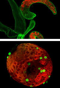 |
| Top: Mutant Drosophila adult testis showing germ cells (red) and cell surface and fusome (green). Bottom: Early stage (third larval instar) wild-type (normal) Drosophila testis showing germ cells (red) and cell surface and pigment cell nuclei (green). Credit: van Doren Lab/JHU |
Abstract:
Seeing something as small as a nanometer—just about three to five atoms wide—requires highly advanced imaging techniques.
Integrated Imaging Center Brings It All into View
Baltimore, MD | Posted on September 17th, 2007Faculty affiliated with Johns Hopkins University's Institute for NanoBioTechnology and researchers from a wide range of disciplines across divisions at JHU will find advanced, comprehensive, and highly precise methods of imaging at the Integrated Imaging Center (IIC) at JHU's Homewood Campus.
Everyone is invited to see firsthand what IIC has to offer at the center's annual open house, co-sponsored by Carl Zeiss and FEICO, for Sept. 21, 2007 from 3 to 6 p.m.
IIC's facilities help scientists and engineers characterize nanomaterials at very small length scale as well as biologists studying complex cellular and subcellular interactions. Microscopy services at IIC also aid developers of medical applications investigate the interface between materials and biological systems and basic biomedical researchers describe nano-sized drug delivery systems.
The center boasts more than $3.5 million worth of state-of-the-art imaging equipment, including one of only two uniquely configured laser scanning microscopes in the United States. The goal of IIC, says center director J. Michael McCaffery, an associate research professor in the Department of Biology, is to provide the Johns Hopkins community, as well as other academic institutions, industry, and government, with resources for both conventional and advanced methods of light and fluorescence microscopy services. IIC's 2,500 square-foot facility is located on the first floor of Dunning Hall.
"As rapid advances have been made in the development of new techniques of fluorescence and electron microscopy, visualization and localization of biomolecules at the light and electron microscope level has become an essential component of any comprehensive study of molecular cell biology," McCaffery says. "This is because light and electron microscopy observations provide detailed information on the distribution, movement, and interaction of biomolecules or proteins within the cell that cannot be obtained by other methods."
To anticipate the changing demands of research in a variety of disciplines, McCaffery says, IIC has added new equipment in recent months. A highlight includes a Zeiss laser scanning microscope (LSM) 510 VIS confocal with a Confocor 3 fluorescence correlation spectroscopy (FCS) module.
"There is only one other system of its kind in the U.S. that combines the FCS with confocal imaging and that is capable of cross-correlation," McCaffery says. "FCS allows for high-resolution spatial and temporal analysis of single biomolecules with respect to diffusion, binding, and enzymatic reactions in vitro and in vivo."
Other new additions include a FEI Tecnai 12 TWIN 120kV high resolution (<2 nm) transmission electron microscope (TEM); a FEI Quanta 200 environmental scanning electron microscope (ESEM), and a dual camera Marianas 4D light microscope (LM) live cell imaging workstation.
The ESEM has proved to be very popular, McCaffery says, because of its ability to "image fully hydrated/wet samples with minimal preparation." In traditional scanning electron microscopy, samples must be dehydrated and coated with a metal prior to imaging. In the ESEM mode, McCaffery explains, "you can use a peltier stage to achieve relative humidity at any temperature or pressure in order to image live samples with minimal surface tension induced drying artifacts."
The center features five suites devoted to specific imaging functions. These include a ultramicrotomy/tissue culture/cell prep room; a wet laboratory; "scanning room," which includes the Quanta ESEM and Typhoon phosphorimager; a transmission electron microscopy suite with two TEMs; and a multifunctional light microscopy suite, which includes the Marians 4D LM, Zeiss LSM 510 VIS confocal with Confocor 3 FCS, LSM 510 META UV confocal, and two Zeiss epifluorescence microscopes. Knowledgeable IIC staff members are available to answer questions or assist with the use of all equipment. Grants from the National Institutes of Health, National Science Foundation, and Howard Hughes Medical Institute help fund the additions to IIC.
The Homewood campus IIC was established in 1998. Additional advanced imaging services have been available at the Montgomery County Campus IIC since 2004.
To learn more about the Integrated Imaging Center at JHU or to schedule an appointment to use the facilities, please go to the IIC Web site: http://www.jhu.edu/~iic/.
To view images from the IIC, click on the following link: http://www.jhu.edu/iic/gallery.htm
####
About Institute for NanoBioTechnology
The Institute for NanoBioTechnology at Johns Hopkins University will revolutionize health care by bringing together internationally renowned expertise in medicine, engineering, the sciences, and public health to create new knowledge and groundbreaking technologies.
INBT programs in research, education, outreach, and technology transfer are designed to foster the next wave of nanobiotechnology innovation.
Approximately 140 faculty are affiliated with INBT and are also members of the following Johns Hopkins institutions: Krieger School of Arts and Sciences, Whiting School of Engineering, School of Medicine, Bloomberg School of Public Health, and Applied Physics Laboratory.
For more information, please click here
Contacts:
* Institute for NanoBioTechnology
214 Maryland Hall
3400 North Charles Street
Baltimore, MD 21218
* Email:
* Phone: (410) 516-3423
* Fax: (410) 516-2355
Copyright © Institute for NanoBioTechnology
If you have a comment, please Contact us.Issuers of news releases, not 7th Wave, Inc. or Nanotechnology Now, are solely responsible for the accuracy of the content.
Bookmark:


| Related News Press |
Academic/Education
![]() Rice University launches Rice Synthetic Biology Institute to improve lives January 12th, 2024
Rice University launches Rice Synthetic Biology Institute to improve lives January 12th, 2024
![]() Multi-institution, $4.6 million NSF grant to fund nanotechnology training September 9th, 2022
Multi-institution, $4.6 million NSF grant to fund nanotechnology training September 9th, 2022
Announcements
![]() NRL charters Navy’s quantum inertial navigation path to reduce drift April 5th, 2024
NRL charters Navy’s quantum inertial navigation path to reduce drift April 5th, 2024
![]() Discovery points path to flash-like memory for storing qubits: Rice find could hasten development of nonvolatile quantum memory April 5th, 2024
Discovery points path to flash-like memory for storing qubits: Rice find could hasten development of nonvolatile quantum memory April 5th, 2024
Tools
![]() Ferroelectrically modulate the Fermi level of graphene oxide to enhance SERS response November 3rd, 2023
Ferroelectrically modulate the Fermi level of graphene oxide to enhance SERS response November 3rd, 2023
![]() The USTC realizes In situ electron paramagnetic resonance spectroscopy using single nanodiamond sensors November 3rd, 2023
The USTC realizes In situ electron paramagnetic resonance spectroscopy using single nanodiamond sensors November 3rd, 2023
Events/Classes
![]() Researchers demonstrate co-propagation of quantum and classical signals: Study shows that quantum encryption can be implemented in existing fiber networks January 20th, 2023
Researchers demonstrate co-propagation of quantum and classical signals: Study shows that quantum encryption can be implemented in existing fiber networks January 20th, 2023
|
|
||
|
|
||
| The latest news from around the world, FREE | ||
|
|
||
|
|
||
| Premium Products | ||
|
|
||
|
Only the news you want to read!
Learn More |
||
|
|
||
|
Full-service, expert consulting
Learn More |
||
|
|
||